Télécharger la présentation
La présentation est en train de télécharger. S'il vous plaît, attendez

1
Longueur télomérique placentaire et retard de croissance intra-utérin
Toutain J (1,2), Prochazkova-Carlotti M (1), Chevret E (1), Cappellen D (1), Merlio JP (1), Horovitz J (3), Saura R (2). (1) Univ. Bordeaux, Histologie et pathologie moléculaire des tumeurs, EA 2406, F Bordeaux, France, (2) CHU Bordeaux, Génétique médicale, F Bordeaux, France, (3) CHU Bordeaux, Gynécologie-obstétrique et médecine fœtale, F Bordeaux, France.
, Prochazkova-Carlotti M (1), Chevret E (1), Cappellen D (1), Merlio JP (1), Horovitz J (3), Saura R (2). (1) Univ. Bordeaux, Histologie et pathologie moléculaire des tumeurs, EA 2406, F Bordeaux, France, (2) CHU Bordeaux, Génétique médicale, F Bordeaux, France, (3) CHU Bordeaux, Gynécologie-obstétrique et médecine fœtale, F Bordeaux, France.")
2
Les télomères

3
Les chromosomes humains
ADN + protéines (histones, non-histones) = chromatine Unité de la chromatine = nucléosome Centromère ADN satellite Kinétochores CENP Télomères (TTAGGG)n Complexe protéique (Segal et al., Nature 2006) (Houben et al., Free Radic Biol Med 2008)
 = chromatine. Unité de la chromatine = nucléosome. Centromère. ADN satellite. Kinétochores. CENP. Télomères. (TTAGGG)n. Complexe protéique. (Segal et al., Nature 2006) (Houben et al., Free Radic Biol Med 2008)")
4
Historique du concept de télomères
HJ Muller (1938) Délétions ou inversions induites par irradiations Mais indétectables aux régions terminales Télomères: telos (fin) et meros (partie) B McClintock (1939) Chromosomes avec cassures double brin Capables de fusionner entre eux Contrairement aux extrémités Structure particulière, capable de stabiliser les chromosomes Telomeres are nucleoprotein structures located at the end of chromosomes. Telomeres consist of repetitive DNA sequences of hexameres « TTAGGG ». The mean telomere length is about of 10 kb, but telomere length closely depends on the tissue studied. Telomeres are essential for chromosome stability. Telomeres especially protect chromosomes from end-to-end fusion or degradation. Telomeres are progressively shortened, especially during mitosis, but telomere length is also influenced by environmental factors such as oxidative stress as hypoxia.
 Délétions ou inversions induites par irradiations. Mais indétectables aux régions terminales. Télomères: telos (fin) et meros (partie) B McClintock (1939) Chromosomes avec cassures double brin. Capables de fusionner entre eux. Contrairement aux extrémités. Structure particulière, capable de stabiliser les chromosomes. Telomeres are nucleoprotein structures located at the end of chromosomes. Telomeres consist of repetitive DNA sequences of hexameres « TTAGGG ». The mean telomere length is about of 10 kb, but telomere length closely depends on the tissue studied. Telomeres are essential for chromosome stability. Telomeres especially protect chromosomes from end-to-end fusion or degradation. Telomeres are progressively shortened, especially during mitosis, but telomere length is also influenced by environmental factors such as oxidative stress as hypoxia.")
5
Les télomères - structure
Structures nucléoprotéiques à l’extrémité des chromosomes Séquences répétitives « TTAGGG » (structure bien conservée) + complexe shelterin Structure double brin et simple brin terminale (~100 nucléotides) « Boucle T » et « boucle D » Longueur télomérique ~10 kb Telomeres are nucleoprotein structures located at the end of chromosomes. Telomeres consist of repetitive DNA sequences of hexameres « TTAGGG ». The mean telomere length is about of 10 kb, but telomere length closely depends on the tissue studied. Telomeres are essential for chromosome stability. Telomeres especially protect chromosomes from end-to-end fusion or degradation. Telomeres are progressively shortened, especially during mitosis, but telomere length is also influenced by environmental factors such as oxidative stress as hypoxia. (d’après Stewart et al., Mutat Res 2012)
 + complexe shelterin. Structure double brin et simple brin terminale (~100 nucléotides) « Boucle T » et « boucle D » Longueur télomérique ~10 kb. Telomeres are nucleoprotein structures located at the end of chromosomes. Telomeres consist of repetitive DNA sequences of hexameres « TTAGGG ». The mean telomere length is about of 10 kb, but telomere length closely depends on the tissue studied. Telomeres are essential for chromosome stability. Telomeres especially protect chromosomes from end-to-end fusion or degradation. Telomeres are progressively shortened, especially during mitosis, but telomere length is also influenced by environmental factors such as oxidative stress as hypoxia. (d’après Stewart et al., Mutat Res 2012)")
6
Longueur télomérique et contraintes réplicatives
Direction de la fourche de réplication 3’ Brin précoce 5’ Hélicase Topoisomérase 5’ ADN polymérase 3’ Extrémité télomérique ARN amorce Brin retardé 5’ Perte longueur télomérique à chaque division Témoin du vieillissement cellulaire

7
Les télomères - fonctions
« Capuchons de protection » « Boucle D » empêche reconnaissance cassures double brin Arrêt du cycle cellulaire Prévention apoptose Prévention phénomènes de dégradation (exonucléases) Prévention phénomènes de fusion des extrémités télomériques (NHEJ) Telomeres are nucleoprotein structures located at the end of chromosomes. Telomeres consist of repetitive DNA sequences of hexameres « TTAGGG ». The mean telomere length is about of 10 kb, but telomere length closely depends on the tissue studied. Telomeres are essential for chromosome stability. Telomeres especially protect chromosomes from end-to-end fusion or degradation. Telomeres are progressively shortened, especially during mitosis, but telomere length is also influenced by environmental factors such as oxidative stress as hypoxia.
 Prévention phénomènes de fusion des extrémités télomériques (NHEJ) Telomeres are nucleoprotein structures located at the end of chromosomes. Telomeres consist of repetitive DNA sequences of hexameres « TTAGGG ». The mean telomere length is about of 10 kb, but telomere length closely depends on the tissue studied. Telomeres are essential for chromosome stability. Telomeres especially protect chromosomes from end-to-end fusion or degradation. Telomeres are progressively shortened, especially during mitosis, but telomere length is also influenced by environmental factors such as oxidative stress as hypoxia.")
8
Longueur télomérique et sénescence
Cellules somatiques, raccourcissement progressif LT A chaque division mitotique Facteurs environnementaux (stress oxydatif) LT réduite → arrêt de la division mitotique (sénescence réplicative) Mécanisme de type suppresseur de tumeur Cellules souches et germinales Potentiel réplicatif élevé Activité télomérase Two words about the enzyme telomerase. This enzyme is able to add telomere repeats at the ends of chromosomes. Telomerase activity is not always detectable. For example, no telomerase activity is detected in somatic cells, but telomerase activity is detected in germinal cells. Telomerase consists of two main subunits: hTERC and hTERT. hTERC subunit is the RNA template necessary to telomerase. hTERC gene is located at locus 3q26. The second subunit is hTERT which is the catalytic subunit of telomerase. hTERT gene is located at locus 5p15. Telomerase activity is normally detected in placentas, but absence of placental telomerase activity has been reported in idiopathic IUGR.
 LT réduite → arrêt de la division mitotique (sénescence réplicative) Mécanisme de type suppresseur de tumeur. Cellules souches et germinales. Potentiel réplicatif élevé. Activité télomérase. Two words about the enzyme telomerase. This enzyme is able to add telomere repeats at the ends of chromosomes. Telomerase activity is not always detectable. For example, no telomerase activity is detected in somatic cells, but telomerase activity is detected in germinal cells. Telomerase consists of two main subunits: hTERC and hTERT. hTERC subunit is the RNA template necessary to telomerase. hTERC gene is located at locus 3q26. The second subunit is hTERT which is the catalytic subunit of telomerase. hTERT gene is located at locus 5p15. Telomerase activity is normally detected in placentas, but absence of placental telomerase activity has been reported in idiopathic IUGR.")
9
L’enzyme télomérase E Blackburn et coll. (1984)
Existence d’une enzyme spécifique : la télomérase (modèle Tetrahymena) Prévient le raccourcissement télomérique physio. en ajoutant des répétitions télomériques aux extrémités Absente des cellules somatiques normales Activée dans cellules souches et germinales Et dans environ 85% des cancers
 Prévient le raccourcissement télomérique physio. en ajoutant des répétitions télomériques aux extrémités. Absente des cellules somatiques normales. Activée dans cellules souches et germinales. Et dans environ 85% des cancers.")
10
L’enzyme télomérase Réverse transcriptase capable d’ajouter des répétions télomériques 2 principales sous-unités: TERT (locus 5p15) TERC (locus 3q26) Two words about the enzyme telomerase. This enzyme is able to add telomere repeats at the ends of chromosomes. Telomerase activity is not always detectable. For example, no telomerase activity is detected in somatic cells, but telomerase activity is detected in germinal cells. Telomerase consists of two main subunits: hTERC and hTERT. hTERC subunit is the RNA template necessary to telomerase. hTERC gene is located at locus 3q26. The second subunit is hTERT which is the catalytic subunit of telomerase. hTERT gene is located at locus 5p15. Telomerase activity is normally detected in placentas, but absence of placental telomerase activity has been reported in idiopathic IUGR. (d’après Artandi et DePinho, Carcinogenesis 2012)
 TERC (locus 3q26) Two words about the enzyme telomerase. This enzyme is able to add telomere repeats at the ends of chromosomes. Telomerase activity is not always detectable. For example, no telomerase activity is detected in somatic cells, but telomerase activity is detected in germinal cells. Telomerase consists of two main subunits: hTERC and hTERT. hTERC subunit is the RNA template necessary to telomerase. hTERC gene is located at locus 3q26. The second subunit is hTERT which is the catalytic subunit of telomerase. hTERT gene is located at locus 5p15. Telomerase activity is normally detected in placentas, but absence of placental telomerase activity has been reported in idiopathic IUGR. (d’après Artandi et DePinho, Carcinogenesis 2012)")
11
Homéostasie de l’oxygène et télomères
Hypoxie → HIF1α → transcription hTERT Stress oxydatif → diminution significative de la longueur télomérique → 8-oxoG RCIU « vasculaire » → stress oxydatif chambre intervilleuse Telomeres are nucleoprotein structures located at the end of chromosomes. Telomeres consist of repetitive DNA sequences of hexameres « TTAGGG ». The mean telomere length is about of 10 kb, but telomere length closely depends on the tissue studied. Telomeres are essential for chromosome stability. Telomeres especially protect chromosomes from end-to-end fusion or degradation. Telomeres are progressively shortened, especially during mitosis, but telomere length is also influenced by environmental factors such as oxidative stress as hypoxia.

12
Etudes portant sur les télomères…

14
Le retard de croissance intra-utérin

15
Le retard de croissance intra-utérin
Définition Jusqu’en 1960 : nouveau-né < 2500 grammes Courbes de poids en fonction de l’âge gestationnel (Lubchenco et al., 1963) Incapacité du fœtus à réaliser son potentiel génétique de croissance Why look at the placental telomere length in pregnancies complicated with intra-uterine growth restriction? Because IUGR is mainly associated with placental insufficiency, and placental insufficiency induces hypoxia. And as we saw earlier, the telomere length is reduced under hypoxia…
 Incapacité du fœtus à réaliser son potentiel génétique de croissance. Why look at the placental telomere length in pregnancies complicated with intra-uterine growth restriction Because IUGR is mainly associated with placental insufficiency, and placental insufficiency induces hypoxia. And as we saw earlier, the telomere length is reduced under hypoxia…")
16
Diagnostic du RCIU A partir de la 20ème SA
Biométries fœtales < 10ème percentile RCIU affirmé PA < 3ème percentile Ou infléchissement courbe de croissance (contrôle à 2 semaines) Why look at the placental telomere length in pregnancies complicated with intra-uterine growth restriction? Because IUGR is mainly associated with placental insufficiency, and placental insufficiency induces hypoxia. And as we saw earlier, the telomere length is reduced under hypoxia… (Conduites pratiques en médecine fœtale, Benachi, 2010)
 Why look at the placental telomere length in pregnancies complicated with intra-uterine growth restriction Because IUGR is mainly associated with placental insufficiency, and placental insufficiency induces hypoxia. And as we saw earlier, the telomere length is reduced under hypoxia… (Conduites pratiques en médecine fœtale, Benachi, 2010)")
17
Conséquences des RCIU Pré-éclampsie (15% des RCIU « vasculaires »),
RCIU associés à une mortalité néonatale et une morbidité infantile et adulte (maladies cardio-vasculaires, obésité, diabète) Problème de santé publique Why look at the placental telomere length in pregnancies complicated with intra-uterine growth restriction? Because IUGR is mainly associated with placental insufficiency, and placental insufficiency induces hypoxia. And as we saw earlier, the telomere length is reduced under hypoxia…
 Problème de santé publique. Why look at the placental telomere length in pregnancies complicated with intra-uterine growth restriction Because IUGR is mainly associated with placental insufficiency, and placental insufficiency induces hypoxia. And as we saw earlier, the telomere length is reduced under hypoxia…")
18
Etiologies du RCIU Fœtales [(cyto)génétiques, infections, GM..] ~25%
Placentaires (anomalies chromosomiques limitées au placenta, anomalies vascularisation utéroplacentaires, anomalies morphologiques..) ~35% Maternelles (toxiques, malnutrition..) ~15% Why look at the placental telomere length in pregnancies complicated with intra-uterine growth restriction? Because IUGR is mainly associated with placental insufficiency, and placental insufficiency induces hypoxia. And as we saw earlier, the telomere length is reduced under hypoxia… (Conduites pratiques en médecine fœtale, Benachi, 2010)
![Etiologies du RCIU Fœtales [(cyto)génétiques, infections, GM..] ~25%](http://slideplayer.fr/slide/1209607/3/images/18/Etiologies+du+RCIU+F%C5%93tales+%5B%28cyto%29g%C3%A9n%C3%A9tiques%2C+infections%2C+GM..%5D+%7E25%25.jpg "Placentaires (anomalies chromosomiques limitées au placenta, anomalies vascularisation utéroplacentaires, anomalies morphologiques..) ~35% Maternelles (toxiques, malnutrition..) ~15% Why look at the placental telomere length in pregnancies complicated with intra-uterine growth restriction Because IUGR is mainly associated with placental insufficiency, and placental insufficiency induces hypoxia. And as we saw earlier, the telomere length is reduced under hypoxia… (Conduites pratiques en médecine fœtale, Benachi, 2010)")
19
Etiologies du RCIU ~25% des RCIU idiopathiques
En majorité insuffisance utéroplacentaire (~60% des RCIU) (Lyall, Placenta 2002) Remodelage réduit des artères spiralées Why look at the placental telomere length in pregnancies complicated with intra-uterine growth restriction? Because IUGR is mainly associated with placental insufficiency, and placental insufficiency induces hypoxia. And as we saw earlier, the telomere length is reduced under hypoxia…
 (Lyall, Placenta 2002) Remodelage réduit des artères spiralées. Why look at the placental telomere length in pregnancies complicated with intra-uterine growth restriction Because IUGR is mainly associated with placental insufficiency, and placental insufficiency induces hypoxia. And as we saw earlier, the telomere length is reduced under hypoxia…")
20
Implantation et développement
Différentiation du trophoblaste Cytotrophoblaste Syncitiotrophoblaste Villosités Ir, IIr et IIIr Réaction déciduale Migration des cellules du trophoblaste extra-villeux, « première vague d’invasion » (1er trimestre, 6ème semaine)
")
21
Implantation et développement
« Deuxième vague d’invasion » au 2ème trimestre (14ème SA) Remodelage des artères spiralées Disparition muscle lisse des artères Flux sanguin utéro-placentaire augmenté Si remodelage imparfait : risque de RCIU +/- signes clinico-biologiques de pré-éclampsie
 Remodelage des artères spiralées. Disparition muscle lisse des artères. Flux sanguin utéro-placentaire augmenté. Si remodelage imparfait : risque de RCIU +/- signes clinico-biologiques de pré-éclampsie.")
22
Etiologies du RCIU (d’après Pijnenborg et al., Placenta 2006)
Why look at the placental telomere length in pregnancies complicated with intra-uterine growth restriction? Because IUGR is mainly associated with placental insufficiency, and placental insufficiency induces hypoxia. And as we saw earlier, the telomere length is reduced under hypoxia… (d’après Pijnenborg et al., Placenta 2006)
")
23
Conséquences au niveau de la chambre intervilleuse
Diminution du débit sanguin maternel (hypoxie) (Biron-Shental et al., AJOG 2010) Ischémie reperfusion (stress oxydatif) (Burton et al., Placenta 2009) Lésions des villosités placentaires (examen histo-pathologique) Why look at the placental telomere length in pregnancies complicated with intra-uterine growth restriction? Because IUGR is mainly associated with placental insufficiency, and placental insufficiency induces hypoxia. And as we saw earlier, the telomere length is reduced under hypoxia…
 (Biron-Shental et al., AJOG 2010) Ischémie reperfusion (stress oxydatif) (Burton et al., Placenta 2009) Lésions des villosités placentaires (examen histo-pathologique) Why look at the placental telomere length in pregnancies complicated with intra-uterine growth restriction Because IUGR is mainly associated with placental insufficiency, and placental insufficiency induces hypoxia. And as we saw earlier, the telomere length is reduced under hypoxia…")
25
Télomères et RCIU

26
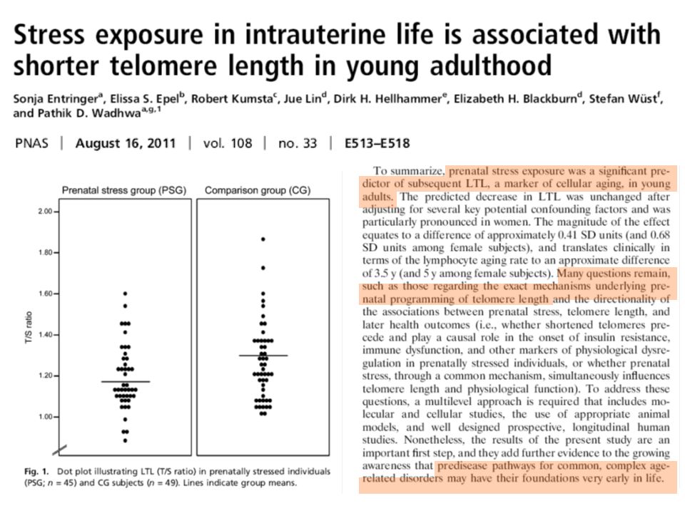
Two recent studies have therefore investigated the placental telomere length at delivery of pregnancies complicated with IUGR. In this first study, placental biopsies were performed at delivery from 27 to 40 weeks. Telomere length was significantly reduced in placenta of pregnancies complicated with IUGR. An association between suppression of telomerase activity and reduced telomere length was also noticed in placentas at delivery of pregnancies complicated with IUGR.

27
A second study confirmed that telomeres were shorter in placentas at delivery of these pregnancies. This study also indicated that telomere length was reduced in placentas at delivery in case of preeclampsia without IUGR. However, no studies have looked to date at telomere length in placentas during ongoing pregnancies at the second and third trimester. This was the main objective of our study. Etudes réalisées au niveau de placentas à terme Longueur télomérique réduite au cours de la grossesse (2ème et 3ème trimestre) ?
")
28
It has also been reported some chromosomal gains of the hTERC telomerase subunit in placentas at delivery of normal pregnancies, while no gains of hTERC were noticed in placentas of pregnancies complicated with IUGR. We wished to assess hTERC and hTERT chromosomal rearrangements, such as gains or losses, in placentas during ongoing pregnancies with and without IUGR at the second and third trimester. This was the secondary objective of our study.

29
Objectifs de l’étude Longueur télomèrique placentaire au cours de grossesses évolutives chez RCIU et contrôles Nombre de copies des loci portant hTERC et hTERT au niveau placentaire

30
Centre de Médecine Fœtale du CHU de Bordeaux
Plus de biopsies de villosités choriales et placentaires depuis 1983 (Saura et coll. Prenat Diagn 2010) Placentocentèse devant tout RCIU inexpliqué : Examen histo-pathologique (Carles et al., Bull Acad Natl Med 2009) Eliminer une ACLP de type 3 Importantes quantités de fragments villositaires At the fetal medicine center of Bordeaux, we have an expertise of early and late CVS, with more than 26,000 CVS performed since 1983. We proposed in case of IUGR to perform the prenatal diagnosis by late CVS rather than by amniocentesis in order to especially eliminate a type 3 confined placental mosaicism. The quantity of chorionic villi obtained in our center allowed us to dispose of an excess of villi to evaluate telomere length and to study rearrangements of hTERC and hTERT loci.
 Placentocentèse devant tout RCIU inexpliqué : Examen histo-pathologique (Carles et al., Bull Acad Natl Med 2009) Eliminer une ACLP de type 3. Importantes quantités de fragments villositaires. At the fetal medicine center of Bordeaux, we have an expertise of early and late CVS, with more than 26,000 CVS performed since We proposed in case of IUGR to perform the prenatal diagnosis by late CVS rather than by amniocentesis in order to especially eliminate a type 3 confined placental mosaicism. The quantity of chorionic villi obtained in our center allowed us to dispose of an excess of villi to evaluate telomere length and to study rearrangements of hTERC and hTERT loci.")
31
Patientes 24 RCIU sévère (cas) et 28 contrôles (HT21, antécédent, âge maternel…) Placentocentèse entre 18 et 37 WA In this monocenter prospective study, 24 patients with idiopathic severe IUGR were included. 28 patients were used as controls. For controls, prenatal diagnosis by late CVS was performed for positive second trimester maternal serum screening, antecedent of chromosomal abnormality, advanced maternal age or isolated ultrasound anomaly. Late CVS were performed between 18 and 37 weeks for cases and controls. We can see in this petri dish the high amount of villi collected by our operator. Routine examinations included interphase FISH for the main aneuploidies and conventional karyotyping. For cases, an histo-pathological examination of placental villi was performed to search for histological signs of placental insufficiency. For this study, excess of villi allowed us to prepare 2 LabtekTM chamber slides for the quantitative FISH technique and 1 flask for the quantitative PCR technique.

32
Caractéristiques des patientes et des grossesses

33
Comment mesurer la longueur télomérique?
Mesure absolue : Southern blot Mesure relative : Quantitative Fluorescent In Situ Hybridization (Q-FISH) Sondes PNA (PanagenTM) lames de culture Microscope automatisé (MetasystemTM) Intensité de fluorescence des sondes télomériques proportionnelle à la longueur des télomères Q-PCR (confirmation) (Cawthon, Nucleic Acid Res 2009) First of all, how to measure telomere length? You can obtain an absolute measurement of telomere length by using a southern blot technique. We have chosen to perform relative measurement of telomere length by a quantitative FISH technique using PNA probes and an automated microscope. Telomere length was supposed to be proportional to the fluorescence intensity of the PNA probes. We confirmed quantitative FISH results by a quantitative PCR technique using specific PCR primers.
 Sondes PNA (PanagenTM) lames de culture. Microscope automatisé (MetasystemTM) Intensité de fluorescence des sondes télomériques proportionnelle à la longueur des télomères. Q-PCR (confirmation) (Cawthon, Nucleic Acid Res 2009) First of all, how to measure telomere length You can obtain an absolute measurement of telomere length by using a southern blot technique. We have chosen to perform relative measurement of telomere length by a quantitative FISH technique using PNA probes and an automated microscope. Telomere length was supposed to be proportional to the fluorescence intensity of the PNA probes. We confirmed quantitative FISH results by a quantitative PCR technique using specific PCR primers.")
34
FISH quantitative - estimation LT
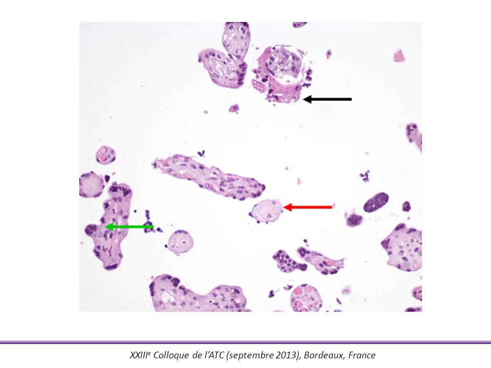
Interphase nuclei (n) 1 2 Fluorescence intensity 1. Villosités placentaires excédentaires, 2 LabtekTM For the quantitative FISH technique, excess of placental villi was cultivated on two LabtekTM chamber slides. Telomere PNA probes were next hybridized on interphase nuclei and the mean intensity of fluorescence was estimated from 1000 interphase nuclei, as illustrated in the figure on the right side. I do not describe here the quantitative PCR technique. 2. Hybridation sondes PNA 3. Distribution intensités de fluorescence télomérique de 1000 noyaux
 Fluorescence intensity. 1. Villosités placentaires excédentaires, 2 LabtekTM. For the quantitative FISH technique, excess of placental villi was cultivated on two LabtekTM chamber slides. Telomere PNA probes were next hybridized on interphase nuclei and the mean intensity of fluorescence was estimated from 1000 interphase nuclei, as illustrated in the figure on the right side. I do not describe here the quantitative PCR technique. 2. Hybridation sondes PNA. 3. Distribution intensités de fluorescence télomérique de 1000 noyaux.")
36
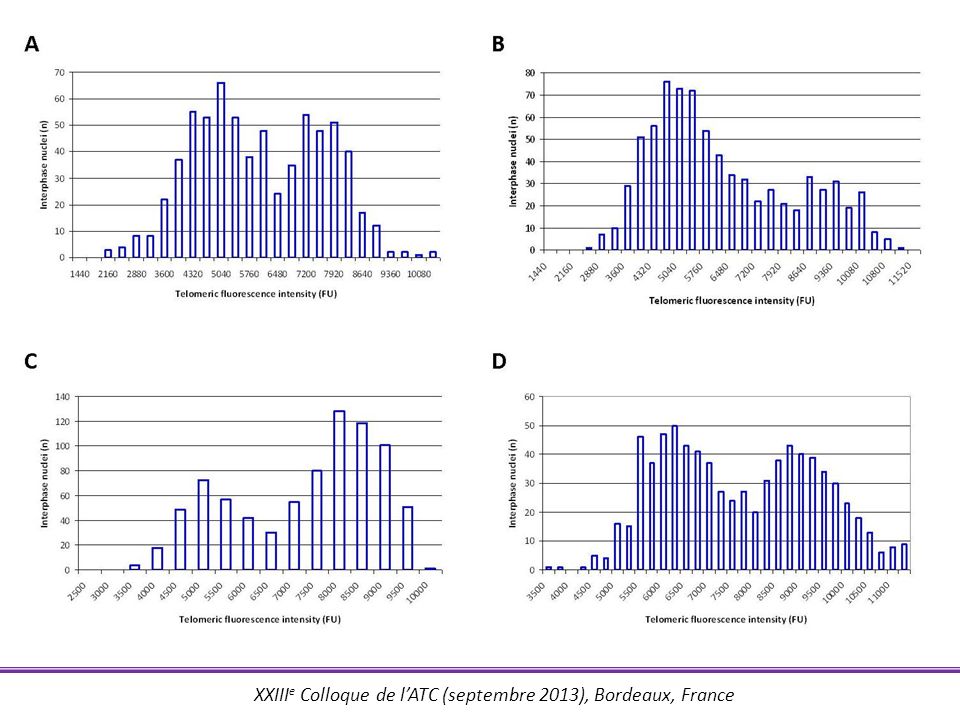
Distribution bimodale après culture
Technique de Cytospin Technique de culture Distribution bimodale après culture

37
Longueur télomérique placentaire et RCIU
Figure A illustrates mean fluorescence intensity of telomere FISH probes according to the term of the placental biopsy for controls (in light grey) and for cases (in dark grey). Figure B indicates that fluorescence intensity was significantly decreased for cases compared to controls. This result indicated that telomere length was significantly reduced in placentas during pregnancies complicated with IUGR.
 and for cases (in dark grey). Figure B indicates that fluorescence intensity was significantly decreased for cases compared to controls. This result indicated that telomere length was significantly reduced in placentas during pregnancies complicated with IUGR.")
38
p < 0,001 p < 0,001 12 controls and 12 cases were randomly selected to perform the quantitative PCR technique. Figure A illustrates that placental telomere length was reduced for the 12 cases with the Q-PCR technique. This result confirmed quantitative FISH results observed for these patients (Figure B). Confirmation en analyse multivariée
. Confirmation en analyse multivariée.")
39
Nombre de copies du locus portant hTERC ?
To study chromosomal rearrangement of hTERC locus, we used a probe at hTERC locus colored in green and a control probe colored in red. Normal hybridization profile is 2 green spots + 2 red spots in each nuclei. An automated microscope captured 300 nuclei and counted the number of green and red spots for each nuclei. hTERC : 3q26.2 (RP11-816J6, vert) Sonde contrôle : 3q13.13 (RP11-16N5, rouge) 300 noyaux interphasiques capturés automatiquement Estimation des gains/pertes du locus portant hTERC
 Sonde contrôle : 3q13.13 (RP11-16N5, rouge) 300 noyaux interphasiques capturés automatiquement. Estimation des gains/pertes du locus portant hTERC.")
40
Nombre de copies du locus portant hTERC ?
Figure A illustrates percentage of nuclei with no chromosomal rearrangement, losses or gains of hTERC locus for controls (light grey) and cases (dark gray). We found no chromosomal rearrangement of hTERC locus for controls and cases. We studied CVS from second and third trimester. hTERC gains were previously described on placentas at delivery. Absence de gain ou de perte du locus portant hTERC Idem pour hTERT
 and cases (dark gray). We found no chromosomal rearrangement of hTERC locus for controls and cases. We studied CVS from second and third trimester. hTERC gains were previously described on placentas at delivery. Absence de gain ou de perte du locus portant hTERC. Idem pour hTERT.")
41
Conclusion / perspectives
LT placentaire réduite entre 18 et 37 SA Non expliquée par copies loci hTERC et hTERT RCIU : stress oxydatif 8-oxoG, sous expression hTERT → absence d’activité télomérase → longueur télomérique placentaire réduite Transcriptome placentaire Marqueurs non invasifs RCIU In summary, we observed that placental telomere length was decreased from the second trimester for pregnancies complicated with IUGR. We did not find chromosomal rearrangement of hTERC locus in placental biopsies of controls and cases. hTERC gains may be therefore a phenomenon of late onset. We think that further studies are needed. Under expression of hTERT causing an absence of telomerase activity, as previously described, may be responsible for short telomere length in placentas for pregnancies complicated with IUGR.

42
Remerciements CHU DE BORDEAUX Université bordeaux segalen
Laboratoire de cytogénétique Pr Robert SAURA Pr Didier LACOMBE Catherine, Céline, Claude, Emeline, Evelyne, Florence, Guillaume, Josiane, Katia, Monique R., Monique V., Natacha, Paulette, Simon, Stéphanie Service d’obstétrique-gynécologie Pr Jacques HOROVITZ (AGOHP) Brigitte, Cathy, Denis , François, Raphaëlle, Sandra, Sara, Suzanne Université bordeaux segalen EA 2406 Histologie et pathologie moléculaire des tumeurs Pr Jean-Philippe MERLIO Pr Pierre DUBUS Alban, Cécile, David, Edith, Elodie, Jacky, Laetitia, Martina, Monique, Olivier, Yamina INSERM U897 Equipe Biostatistique Pr Daniel COMMENGES Ana
 Brigitte, Cathy, Denis , François, Raphaëlle, Sandra, Sara, Suzanne. Université bordeaux segalen. EA 2406 Histologie et pathologie moléculaire des tumeurs. Pr Jean-Philippe MERLIO. Pr Pierre DUBUS. Alban, Cécile, David, Edith, Elodie, Jacky, Laetitia, Martina, Monique, Olivier, Yamina. INSERM U897 Equipe Biostatistique. Pr Daniel COMMENGES. Ana.")
43
Longueur télomérique et stress oxydatif

Présentations similaires
 ET MALADIES GENETIQUES>")


>")


>")


>")
