Télécharger la présentation
La présentation est en train de télécharger. S'il vous plaît, attendez

1
Capacité de gérontologie clinique Vendredi 29 avril 2005
LE LUPUS DU SUJET AGE Dr Gil Helder Praticien Hospitalier Service de Médecine Interne. Besançon.

2
Données épidémiologiques
15 à 50 cas/ hab Fréquence dans la race noire et asiatique Age moyen de diagnostic : environ 30 ans Délai moyen de diagnostic : 1 à 2 ans Prédominance féminine , sex ratio à 13,2 mais diminue avec l'âge Lupus et sujet âgé... Au delà de 50 ans sex ratio à 2,6 En france : 6 à 9 % Et jusqu'à 20 % (Danemark) Peu d'études au delà de 65 ans...
 Peu d études au delà de 65 ans...")
3
Rev Med Interne. 2003 May;24(5):288-94.
[Systemic lupus erythematosus with disease onset after age 65] Gaujard S, Broussolle C, Cathebras P, Dupond JL, Massot C, Ninet J, Perrot H, Durand DV, Rousset H. Hopital geriatrique Antoine-Charial, centre hospitalier universitaire de Lyon, 40, avenue de la Table-de-Pierre, Francheville, France. OBJECTIVES: Systemic lupus erythematosus with disease-onset in the elderly has rarely been studied (only one report about 21 patients with disease onset at 65 and older). Is the management of this pathology modified in this population? METHODS: Seventeen hospitalised cases of lupus patients with disease onset at 65 or older are retrospectively reported between 1988 and The results are compared with those of younger subjects. RESULTS: The female to male ratio is Mean age at disease onset is /- 3.5 years. Mean duration of follow-up is 3.5 +/- 2.4 years. Main initial symptoms are: deterioration of general status (41%), arthritis (35%), cutaneous manifestations (35%), thrombo-embolism (24%) and pleuritis (18%). Malar rash is uncommon (12%). Nephropathy is never a revealing symptom and is rarely serious during the disease's evolution. Like in neurologic manifestations, the etiology has to be discussed in relation to associated co-morbidities. Concerning haematologic features, lymphopenia is found in 82% of the cases with a questionable specificity. Antinuclear antibodies are constant, anti-dsDNA antibodies are found in 82% of the cases, antibodies to extractable nuclear antigens in 50%, and anticoagulant circulating activity in 59%. Prognosis is difficult to assess in such a limited series but 5-years survival probability is 83%. Glucocorticoid lead to 50% of major complications. CONCLUSIONS: This study focuses on the particular initial manifestations of systemic lupus erythematosus in the elderly (deterioration of general status, thrombosis, unusual cutaneous symptoms), and on the specificity of differential diagnosis and treatment. PMID: [PubMed - indexed for MEDLINE]
. Is the management of this pathology modified in this population METHODS: Seventeen hospitalised cases of lupus patients with disease onset at 65 or older are retrospectively reported between 1988 and The results are compared with those of younger subjects. RESULTS: The female to male ratio is Mean age at disease onset is /- 3.5 years. Mean duration of follow-up is 3.5 +/- 2.4 years. Main initial symptoms are: deterioration of general status (41%), arthritis (35%), cutaneous manifestations (35%), thrombo-embolism (24%) and pleuritis (18%). Malar rash is uncommon (12%). Nephropathy is never a revealing symptom and is rarely serious during the disease s evolution. Like in neurologic manifestations, the etiology has to be discussed in relation to associated co-morbidities. Concerning haematologic features, lymphopenia is found in 82% of the cases with a questionable specificity. Antinuclear antibodies are constant, anti-dsDNA antibodies are found in 82% of the cases, antibodies to extractable nuclear antigens in 50%, and anticoagulant circulating activity in 59%. Prognosis is difficult to assess in such a limited series but 5-years survival probability is 83%. Glucocorticoid lead to 50% of major complications. CONCLUSIONS: This study focuses on the particular initial manifestations of systemic lupus erythematosus in the elderly (deterioration of general status, thrombosis, unusual cutaneous symptoms), and on the specificity of differential diagnosis and treatment. PMID: [PubMed - indexed for MEDLINE]")
4
Lupus. 2000;9(2):96-100. Related Articles, Books, LinkOut
Click here to read The clinical features and prognosis of lupus with disease onset at age 65 and older. Pu SJ, Luo SF, Wu YJ, Cheng HS, Ho HH. Department of Internal Medicine, Chang Gung Memorial Hospital, Tao-Yuan, Taiwan. Systemic lupus erythematosus (SLE) in the elderly is uncommon and rarely reported with disease onset at age 65 and older. The aim of this study is to retrospectively analyze the influence of age at disease onset on the clinical features and prognosis of SLE. From 1988 to 1998, we encountered 21 lupus patients with disease onset at age 65 and older (all are included in group A). For comparison, 21 lupus patients with disease onset between years of age (group B) and 152 lupus patients with disease onset before 50 years of age (group C) were obtained by a simple random sampling method from the hospital registry. Clinical features as included in the 1982 ARA revised criteria for classification of SLE and survival rate were analyzed and compared among these three groups. Group A had a smaller female to male ratio, longer duration from disease onset to diagnosis, less malar rash, more discoid lupus, and shorter survival rate that group C. There was no statistically significant difference in clinical features and survival between groups A and B, as well as between female and male patients of these two groups. The main cause of death in group A was septic shock. In conclusion, the clinical features and prognosis of SLE were influenced by the age at disease onset. However, clinical features and prognosis of SLE were similar in both late-onset lupus groups. PMID: [PubMed - indexed for MEDLINE]
 in the elderly is uncommon and rarely reported with disease onset at age 65 and older. The aim of this study is to retrospectively analyze the influence of age at disease onset on the clinical features and prognosis of SLE. From 1988 to 1998, we encountered 21 lupus patients with disease onset at age 65 and older (all are included in group A). For comparison, 21 lupus patients with disease onset between years of age (group B) and 152 lupus patients with disease onset before 50 years of age (group C) were obtained by a simple random sampling method from the hospital registry. Clinical features as included in the 1982 ARA revised criteria for classification of SLE and survival rate were analyzed and compared among these three groups. Group A had a smaller female to male ratio, longer duration from disease onset to diagnosis, less malar rash, more discoid lupus, and shorter survival rate that group C. There was no statistically significant difference in clinical features and survival between groups A and B, as well as between female and male patients of these two groups. The main cause of death in group A was septic shock. In conclusion, the clinical features and prognosis of SLE were influenced by the age at disease onset. However, clinical features and prognosis of SLE were similar in both late-onset lupus groups. PMID: [PubMed - indexed for MEDLINE]")
5
Medicine (Baltimore). 1993 Mar;72(2):113-24.
Systemic lupus erythematosus: clinical and immunologic patterns of disease expression in a cohort of 1,000 patients. The European Working Party on Systemic Lupus Erythematosus. Cervera R, Khamashta MA, Font J, Sebastiani GD, Gil A, Lavilla P, Domenech I, Aydintug AO, Jedryka-Goral A, de Ramon E, et al. Department of Internal Medicine, Hospital Clinic, Barcelona, Spain. In the present study we have analyzed the prevalence and characteristics of the most relevant clinical and immunologic features in 1,000 patients with SLE. Several differences in the expression of the disease have been observed in relation to the patients' age at onset, sex, and autoantibody serology. The childhood-onset patients more often had malar rashes (55% vs 39%) and nephropathy (28% vs 15%) as presenting manifestations. During the evolution of the disease, these patients had an increased prevalence only of malar rash (79% vs 56%) and a lower prevalence of rheumatoid factor (6% vs 19%). The older-onset patients (age 50 or older) less often showed malar rash (21% vs 42%), arthritis (52% vs 71%), and nephropathy (3% vs 17%) as the first symptom. During the evolution of their disease, these patients had a decreased prevalence of malar rash (33% vs 60%), photosensitivity (29% vs 47%), arthritis (73% vs 85%), nephropathy (22% vs 41%), thrombosis (4% vs 15%), and anti-La antibodies (6% vs 20%), but an increased prevalence of sicca syndrome (33% vs 15%). Males more often had serositis (28% vs 16%) as a first symptom, but they presented with a lower prevalence of arthritis (74% vs 85%) during the evolution of the disease. The presence of ANA, a high titer of anti-dsDNA, rheumatoid factor, anti-ENA, and antiphospholipid antibodies also distinguished additional homogeneous SLE subsets of clinical significance.
 and nephropathy (28% vs 15%) as presenting manifestations. During the evolution of the disease, these patients had an increased prevalence only of malar rash (79% vs 56%) and a lower prevalence of rheumatoid factor (6% vs 19%). The older-onset patients (age 50 or older) less often showed malar rash (21% vs 42%), arthritis (52% vs 71%), and nephropathy (3% vs 17%) as the first symptom. During the evolution of their disease, these patients had a decreased prevalence of malar rash (33% vs 60%), photosensitivity (29% vs 47%), arthritis (73% vs 85%), nephropathy (22% vs 41%), thrombosis (4% vs 15%), and anti-La antibodies (6% vs 20%), but an increased prevalence of sicca syndrome (33% vs 15%). Males more often had serositis (28% vs 16%) as a first symptom, but they presented with a lower prevalence of arthritis (74% vs 85%) during the evolution of the disease. The presence of ANA, a high titer of anti-dsDNA, rheumatoid factor, anti-ENA, and antiphospholipid antibodies also distinguished additional homogeneous SLE subsets of clinical significance.")
6
Medicine (Baltimore). 2004 Nov;83(6):348-59.
Late-onset systemic lupus erythematosus: a personal series of 47 patients and pooled analysis of 714 cases in the literature. Boddaert J, Huong du LT, Amoura Z, Wechsler B, Godeau P, Piette JC. Department of Internal Medicine, Groupe hospitalier Pitie-Salpetriere, Paris, France. Systemic lupus erythematosus (SLE) is uncommon after the age of 50 years, and studies of elderly patients with SLE are scarce. We conducted the current study to analyze characteristics and outcome of patients with late-onset SLE in a French tertiary referral center, and to compare them with those of younger patients with SLE. From 1980 to 2000, 47 patients were identified as having late-onset SLE, defined as SLE diagnosed at or over the age of 50 years. These patients were compared with a group of 114 randomly selected patients aged younger than 50 years at SLE diagnosis. We compared clinical characteristics, laboratory data, therapy, and course.The female to male ratio was smaller in the late-onset SLE group (p = ). Some manifestations occurred less frequently in late-onset SLE: arthritis (p = 0.009), malar rash (p = 0.013), and nephropathy (p = 0.009). High-dose corticosteroids (p = ) and immunosuppressive drugs (p = 0.006) were less commonly used in the elderly. Deaths occurred more frequently in late-onset SLE (p = 0.019), with a 10-year survival rate of 71% versus 95% in early-onset SLE (p < 0.01). In patients with late-onset SLE, causes of death were usually unrelated to SLE.Analysis of pooled data from the literature, based on 714 old and 4700 young SLE patients, confirmed that late-onset SLE was characterized by a smaller female to male ratio (4.4:1 vs. 10.6:1; p = 3.10); a higher occurrence of serositis (36.7% vs. 28.6%; p = 7.10) and pulmonary involvement (21.2% vs. 11.3%; p = 6.10); and a lower occurrence of malar rash (31.1% vs. 62.4%; p = 10), photosensitivity (26.2% vs. 38.2%; p = 6.10), purpura/cutaneous vasculitis (13.4% vs. 25.9%; p = 9.10), alopecia/hair loss (24% vs. 44.9%; p = 3.10), Raynaud phenomenon (24.8% vs. 37.2%; p = 3.10), neuropsychiatric manifestations (15.3% vs. 20.2%; p = 0.025), lymphadenopathy (9.1% vs. 19.6%; p = 2.10), nephrotic syndrome (8.1% vs. 24.3%; p = 0.015), and nephritis (28.6% vs. 42.7%; p = 2.10). Regarding laboratory features, rheumatoid factor positivity was more frequent (32.7% vs. 20.1%; p = 3.10), whereas anti-RNP positivity (10.4% vs. 20.9%; p = 9.10), anti-Sm positivity (9.1% vs. 17.1%; p = 0.001), and a low CH50 complement fraction (45% vs. 64.9%; p = 0.002) were less frequent in old compared with young SLE patients.In conclusion, the clinical pattern of late-onset SLE is characterized by a lower disease severity. The reduced survival observed in this group seems to result mainly from the consequences of aging.
 is uncommon after the age of 50 years, and studies of elderly patients with SLE are scarce. We conducted the current study to analyze characteristics and outcome of patients with late-onset SLE in a French tertiary referral center, and to compare them with those of younger patients with SLE. From 1980 to 2000, 47 patients were identified as having late-onset SLE, defined as SLE diagnosed at or over the age of 50 years. These patients were compared with a group of 114 randomly selected patients aged younger than 50 years at SLE diagnosis. We compared clinical characteristics, laboratory data, therapy, and course.The female to male ratio was smaller in the late-onset SLE group (p = ). Some manifestations occurred less frequently in late-onset SLE: arthritis (p = 0.009), malar rash (p = 0.013), and nephropathy (p = 0.009). High-dose corticosteroids (p = ) and immunosuppressive drugs (p = 0.006) were less commonly used in the elderly. Deaths occurred more frequently in late-onset SLE (p = 0.019), with a 10-year survival rate of 71% versus 95% in early-onset SLE (p < 0.01). In patients with late-onset SLE, causes of death were usually unrelated to SLE.Analysis of pooled data from the literature, based on 714 old and 4700 young SLE patients, confirmed that late-onset SLE was characterized by a smaller female to male ratio (4.4:1 vs. 10.6:1; p = 3.10); a higher occurrence of serositis (36.7% vs. 28.6%; p = 7.10) and pulmonary involvement (21.2% vs. 11.3%; p = 6.10); and a lower occurrence of malar rash (31.1% vs. 62.4%; p = 10), photosensitivity (26.2% vs. 38.2%; p = 6.10), purpura/cutaneous vasculitis (13.4% vs. 25.9%; p = 9.10), alopecia/hair loss (24% vs. 44.9%; p = 3.10), Raynaud phenomenon (24.8% vs. 37.2%; p = 3.10), neuropsychiatric manifestations (15.3% vs. 20.2%; p = 0.025), lymphadenopathy (9.1% vs. 19.6%; p = 2.10), nephrotic syndrome (8.1% vs. 24.3%; p = 0.015), and nephritis (28.6% vs. 42.7%; p = 2.10). Regarding laboratory features, rheumatoid factor positivity was more frequent (32.7% vs. 20.1%; p = 3.10), whereas anti-RNP positivity (10.4% vs. 20.9%; p = 9.10), anti-Sm positivity (9.1% vs. 17.1%; p = 0.001), and a low CH50 complement fraction (45% vs. 64.9%; p = 0.002) were less frequent in old compared with young SLE patients.In conclusion, the clinical pattern of late-onset SLE is characterized by a lower disease severity. The reduced survival observed in this group seems to result mainly from the consequences of aging.")
7
Circonstances de découverte
Cas typique : atteinte multiviscérale Plus souvent : Polyarthrite non destructrice Pleuropéricardite récidivante Néphropathie glomérulaire isolée PTI, AHAI... Sujet âgé : Polyarthrite 42 % Pleurésie 15 % Péricardite 13 % Rash malaire 10%

8
critères de l'American College of Rheumatology (1982)
Eruption malaire en aile de papillon Eruption de lupus discoïde Photosensibilité Ulcérations buccales ou naso-pharyngées Polyarthrite non érosive Pleurésie ou péricardite Atteinte rénale (protéinurie>0,5g/j, cylindres urinaires) Atteinte neurologique : convulsion, psychose Atteinte hématologique : anémie hémolytique avec réticulocytose ou leucopénie < ou lymphopénie < 1500 ou thrombopénie < Désordres immunologiques : présence de cellules LE ou Ac anti-DNA natif ou anti- Sm ou fausse sérologie syphilitique Présence de facteurs anti-nucléaires à un titre anormal en l'absence de médicament inducteur Nécessite 4 critères simultanés ou successifs (spécificité et sensibilité > 90 %)
 Atteinte neurologique : convulsion, psychose. Atteinte hématologique : anémie hémolytique avec réticulocytose ou leucopénie < 4000 ou lymphopénie < 1500 ou thrombopénie < Désordres immunologiques : présence de cellules LE ou Ac anti-DNA natif ou anti- Sm ou fausse sérologie syphilitique. Présence de facteurs anti-nucléaires à un titre anormal en l absence de médicament inducteur. Nécessite 4 critères simultanés ou successifs. (spécificité et sensibilité > 90 %)")
9
Signes cliniques classiques (1) :
Signes généraux non spécifiques Atteintes cutanées : Forme aigüe (rare) : oedème érythémateux du visage Vespertilio Lupus discoïde (chronique, atrophie centrale), alopécie Lucite Vascularite (gravité) Autres : angiooedème, bulles, ulcérations muqueuses... Sujet âgé : Rash malaire moins fréquent (25 % vs 42 %) Photosensibilité : études contractoires
 : oedème érythémateux du visage. Vespertilio. Lupus discoïde (chronique, atrophie centrale), alopécie. Lucite. Vascularite (gravité) Autres : angiooedème, bulles, ulcérations muqueuses... Sujet âgé : Rash malaire moins fréquent (25 % vs 42 %) Photosensibilité : études contractoires.")
10
Penser aux anti-phospholipides Attention aux livédos de stase…

11
Ulcération secondaire à un anti-phospholipide

12
Signes cliniques classiques (2) :
Atteintes articulaires et musculaires : souvent inaugurales Arthralgies migratrices Polyarthrites bilat et symétriques (sub aïgues) Polyarthrites chroniques « Rhumatisme de Jaccoud » (destruction ligamentaire et capsulaire) Ostéonécrose aseptique Myalgies et myosites Sujet âgé : arthralgies moins fréquentes en cours d'évolution
 Polyarthrites chroniques « Rhumatisme de Jaccoud » (destruction ligamentaire et capsulaire) Ostéonécrose aseptique. Myalgies et myosites. Sujet âgé : arthralgies moins fréquentes en cours d évolution.")
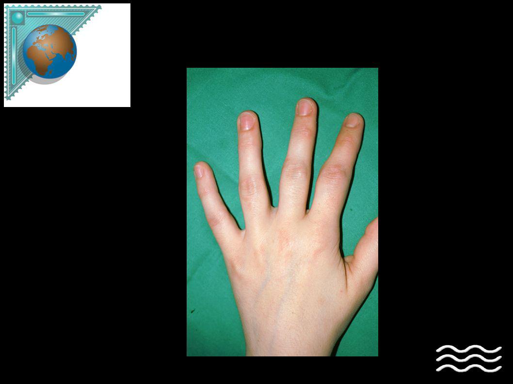
14
polyarthrite non destructrice

15
Atteintes cardio-vasculaires
Raynaud : 30 % HTA, en général secondaire Péricardite +/- pleurésie Sérofibrineuse et corticosensible frottement rare, parfois asymptômatique Myocardite interstitielle penser : SAPL et myocardite ischémique Endocardite de Libman Sachs / surtout si SAPL souffle diastolique Insuffisance coronarienne TVP : 10 à 20 %. SAPL +++ Sujet âgé : Même fréquence de péricardite, plus d'HTA Moindre fréquence du syndrome de Raynaud

16
Atteintes pleuro-pulmonaires
Pleurésie séro-fibrineuse fq et corticosensible Pneumopathie : rare opacités migratrices et nodulaires, basales HTAP : rare / SAPL Hémorragie alvéolaire, atéléctasie des coupoles... Sujet âgé : fréquence des pleurésies (31 vs 18 %)
")
17
Atteintes rénales Facteur pronostique Fréquentes Souvent initiales
Gravité non corrélée à la gravité apparente de la maladie Particularités chez le sujet âgé : moins fréquentes (20 % vs 51 %) protéinurie > 1 g : 17 % vs 38 % insuf rénale : 4 % vs 15 % GM proliférative (classe III et IV)
 protéinurie > 1 g : 17 % vs 38 % insuf rénale : 4 % vs 15 % GM proliférative (classe III et IV)")
18
Atteintes neuro-psychiatriques
40 % au cours de la maladie Centrales : Comitialité et signes extra-pyramidaux Signes focaux : penser SAPL Rares : céphalées, neuropathies périphériques, méningites lymphocytaires Psychiatriques : fq et polymorphes Confusion ou délire aigu Dépression, psychose, désorientation LCR : normal ou lymphocytaire IRM : aspect de microvascularite Sujet âgé : tout est possible difficultés d'interpréter les signes neurologiques...

19
Atteintes rares Formes oculaires : Formes digestives :
Rétinite dysorique Rares : névrite optique, occlusion vx rétiniens Formes digestives : Hémorragie : iatrogène Pancréas : rare et pronostique Pas d'atteinte hépatique spécifique

20
Biologie « conventionnelle » Pas de différence avec les sujets jeunes
Dysglobulinémie avec élévation des IgG Peu inflammatoire (sauf épcht des séreuses) Leucopénie (neutropénie et/ou lymphopénie) Anémie hémolytique Thrombopénie auto-immune
 Leucopénie (neutropénie et/ou lymphopénie) Anémie hémolytique. Thrombopénie auto-immune.")
21
Biologie « spécifique » (1)
AAN : 95 % de positivité DNA natifs (marquage homogène) : 60 à 80 % de positivité Ag solubles (marquage moucheté) : Sm : spécifique, 30 % RNP : 40 % SSA : 30 % , svt négatif en DNA SSB : Sjogren associé Anti-coagulants circulants Sujets âgés : moindre fréquence des Ag solubles
 : 60 à 80 % de positivité. Ag solubles (marquage moucheté) : Sm : spécifique, 30 % RNP : 40 % SSA : 30 % , svt négatif en DNA. SSB : Sjogren associé. Anti-coagulants circulants. Sujets âgés : moindre fréquence. des Ag solubles.")
22
Biologie « spécifique » (2)
“Facteurs rhumatoïdes” : 20 % Coombs direct : 50 % des cas Complément : consommé dans 50 % des cas C4 et C3 : témoins d'activation de la maladie Intérêt dans l'atteinte rénale Sujet âgé : consommation du complément : moins fréquente plus souvent : présence de facteurs rhumatoïdes

23
Le lupus a-t-il une évolution spécifique chez le sujet âgé ?
Evolution similaire jusqu'à 4 ans après le diagnostic Probabilité de survie (medicine 2004) : À 5 ans : 84 % (vs 95 %) À 10 ans : 71 % (vs 95 %) À 15 ans : 59 % (vs 92 %) Contraste avec la moindre sévérité clinique apparente D'autres études ne montrent pas de différences mais : Critères diagnostiques non réunis Utilisation moindre des immunosuppresseurs
 : À 5 ans : 84 % (vs 95 %) À 10 ans : 71 % (vs 95 %) À 15 ans : 59 % (vs 92 %) Contraste avec la moindre sévérité clinique apparente. D autres études ne montrent pas de différences mais : Critères diagnostiques non réunis. Utilisation moindre des immunosuppresseurs.")
24
FDR classique + corticoides + antiphospholipides + inflammation
Les causes de mortalité du sujet âgé porteur d'un lupus ? Cervera, medicine 2003,72:113 Causes de décès : 26,5 % : lupus 26,5 % : thromboses 25 % : infections Faible mortalité par néoplasie : 2,3 % vs 6% Risque cardiovasculaire augmenté : FDR classique + corticoides + antiphospholipides + inflammation

25
Y-a-t-il un traitement spécifique du lupus chez le sujet âgé ?
Théoriquement : non !

26
Les données de la litterature analyse poolée (sujets âgés vs sujets jeunes)
Clinique SLE âgé (n=714) SLE jeune (n=4700) sex ratio ,4/ ,6/1 rash malaire % % photosensilité % % vascularite cutanée % % alopécie % % arthrite % % Raynaud % % séreuse % % poumon % % neuropsy % % hépatique % 3,5 % adénopathies % % Σ néphrotique % %
 SLE jeune (n=4700) sex ratio 4,4/1 10,6/1. rash malaire 31 % 62 % photosensilité 26 % 38 % vascularite cutanée 13 % 26 % alopécie 24 % 45 % arthrite 66 % 71 % Raynaud 25 % 37 % séreuse 36 % 28 % poumon 21 % 11 % neuropsy 15 % 20 % hépatique 15 % 3,5 % adénopathies 9 % 19 % Σ néphrotique 8 % 24 %")
27
Les données de la litterature analyse poolée (sujets âgés vs sujets jeunes)
Biologie SLE âgé (n=714) SLE jeune (n=4700) Facteurs rhumatoïdes % % baisse du complément % % faux BW % % anti-SM % % anti-RNP % % thérapeutiques corticoïdes % % immunosuppresseurs % %
 SLE jeune (n=4700) Facteurs rhumatoïdes 32 % 20 % baisse du complément 41 % 56 % faux BW 11 % 17 % anti-SM 9 % 17 % anti-RNP 10 % 21 % thérapeutiques. corticoïdes 78 % 85 % immunosuppresseurs 25 % 37 %")
28
Spécificités du diagnostic de lupus au delà de 65 ans
Co-morbidités associées Difficultés à analyser les signes articulaires Intérêt de la biopsie en cas d'insuffisance rénale Interprétation des symptômes neurologiques Fréquence de la lymphopénie Fréquence des auto-anticorps chez le sujet âgé...y compris les anti-coagulants circulants

29
Comparaison lupus après 65 ans et sujets jeunes...
Sex ratio (influence hormonale) Signes articulaires plus fréquents au début...moins fréquents en cours d'évolution Manifestations cutanées sub-aigües moins fréquentes Faible taux de glomérulonéphrite lupique Complications iatrogènes (corticothérapie) Fréquence de l'interruption des anti-paludéens Pas de complication infectieuse ou hématologique majeure sous azathioprine ou ciclophosphamide
 Signes articulaires plus fréquents au début...moins fréquents en cours d évolution. Manifestations cutanées sub-aigües moins fréquentes. Faible taux de glomérulonéphrite lupique. Complications iatrogènes (corticothérapie) Fréquence de l interruption des anti-paludéens. Pas de complication infectieuse ou hématologique majeure sous azathioprine ou ciclophosphamide.")
30
...et entre 50-65 ans par rapport à plus de 65 ans
Peu de différence Sauf dans la présentation initiale : Altération de l'état général Thromboses plus fréquentes

31
Conclusions LED : diagnostic sous estimé
Signes cliniques atypiques (AEG, thrombose, peau, articulations) Le piège de la pleurésie Interprétation des auto-anticorps ?...la clinique !!! Pas de “bricolage thérapeutique”
 Le piège de la pleurésie. Interprétation des auto-anticorps ...la clinique !!! Pas de bricolage thérapeutique")
32
Quelques questions …. Principales manifestations cliniques inaugurales du LED du sujet âgé ? Quelles sont les anomalies biologiques « non immunologiques » observées au cours du LED ? Causes de décès des sujets lupiques âgés Citez les particularités du LED au-delà de 65 ans

Présentations similaires


>")

![[number 1-100] There is no rule to the way to remember the names for the numbers 1 to 10 in French so we recommend that you simply practice!](/1/172873/big_thumb.jpg "[number 1-100] There is no rule to the way to remember the names for the numbers 1 to 10 in French so we recommend that you simply practice!>")


 Michel Tenenhaus.>")