Télécharger la présentation
La présentation est en train de télécharger. S'il vous plaît, attendez

1
Paroi bactérienne Schéma de la paroi d'une bactérie à Gram négatif.
Schéma de la paroi d'une bactérie à Gram positif. Paroi bactérienne

2
Structure schématique du peptidoglycane.

3
Bactéries à Gram positif Bactéries à Gram négatif
Comparaison de la paroi des bactéries à Gram positif et des bactéries à Gram négatif. Bactéries à Gram positif Bactéries à Gram négatif Aspect en microscopie électronique Une couche épaisse et amorphe. Deux couches séparées par un espace clair. Présence d'une membrane externe Non Oui Présence d'un espace périplasmique Peptidoglycane Épais (10 à 80 nm), représente 40 p. cent du poids sec, détermine la morphologie bactérienne. Mince (2 à 6 nm), représente moins de 10 p. cent du poids sec, détermine la morphologie bactérienne. Acides téchoïques Présents Absents Présence de protéines Possible : liaisons covalentes avec le peptidoglycane, rôle éventuel dans le pouvoir pathogène, rôle éventuel dans l'antigénicité spécifique. Fréquente Présence de polysaccharides Possible : antigènes spécifiques de groupe pour certaines espèces Possible Lipopolysaccharides
, représente 40 p. cent du poids sec, détermine la morphologie bactérienne. Mince (2 à 6 nm), représente moins de 10 p. cent du poids sec, détermine la morphologie bactérienne. Acides téchoïques. Présents. Absents. Présence de protéines. Possible : liaisons covalentes avec le peptidoglycane, rôle éventuel dans le pouvoir pathogène, rôle éventuel dans l antigénicité spécifique. Fréquente. Présence de polysaccharides. Possible : antigènes spécifiques de groupe pour certaines espèces. Possible. Lipopolysaccharides.")
4
Schéma général de la cellule (cellule eucaryote animale= 50 µm)
Centrioles Réticulum endoplasmique Lysosomes Peroxysomes Mitochondrie Noyau avec le nucléole(zone sombre) Appareil de Golgi Microfilaments (cytosquelette) Cytoplasme Microtubules (cytosquelette) Ribosomes
 Appareil de Golgi. Microfilaments (cytosquelette) Cytoplasme. Microtubules (cytosquelette) Ribosomes.")
5
Schéma général de la cellule (cellule eucaryote végétale = 200 µm)
Microfilaments (cytosquelette) Ribosomes Cytoplasme Appareil de Golgi Vacuole Reticulum endoplasmique Peroxysomes Microtubules (cytosquelette) Lysosomes Chloroplastes Mitochondries Paroi extra-cellulaire
 Ribosomes. Cytoplasme. Appareil de Golgi. Vacuole. Reticulum endoplasmique. Peroxysomes. Microtubules (cytosquelette) Lysosomes. Chloroplastes. Mitochondries. Paroi extra-cellulaire.")
6
Paroi d’une cellule végétale
Paroi épidermique de soja en croissance La structure de la paroi qui vient d'être secrétée (à l'intérieur, contre la membrane plasmique) est différente de celle de la paroi externe plus agée et qui a donc subie une croissance antérieure (à l'extérieur).
 est différente de celle de la paroi externe plus agée et qui a donc subie une croissance antérieure (à l extérieur).")
7
Membrane plasmique observée au MET

20
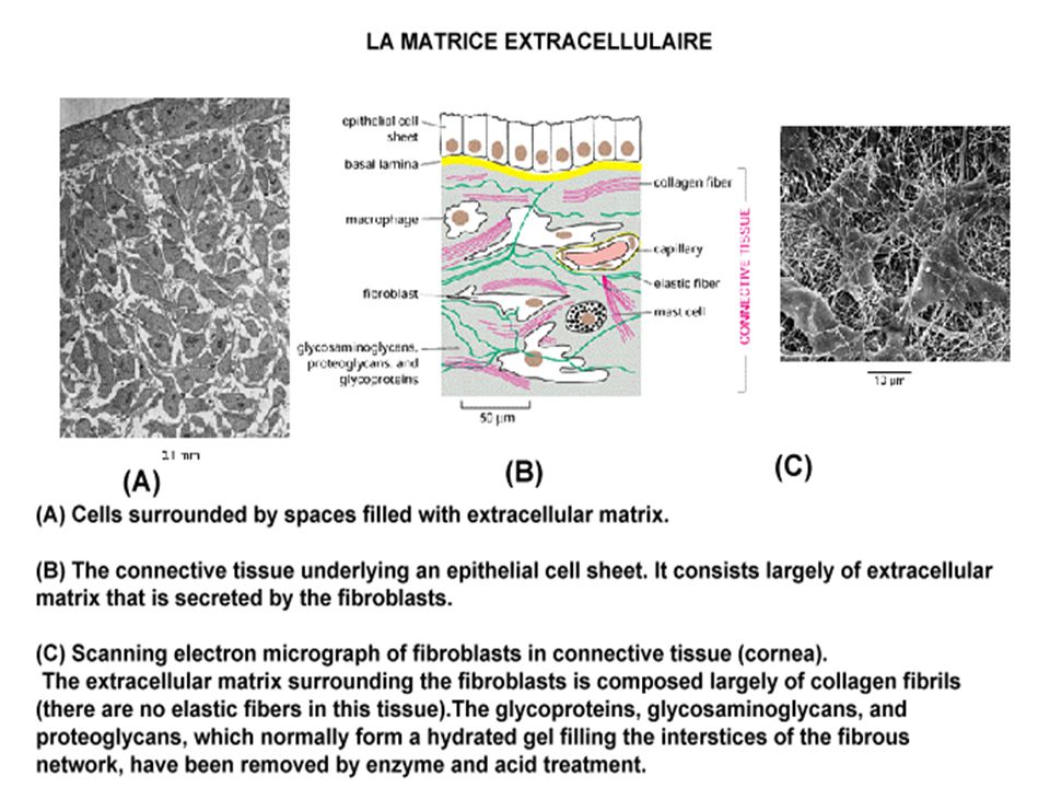
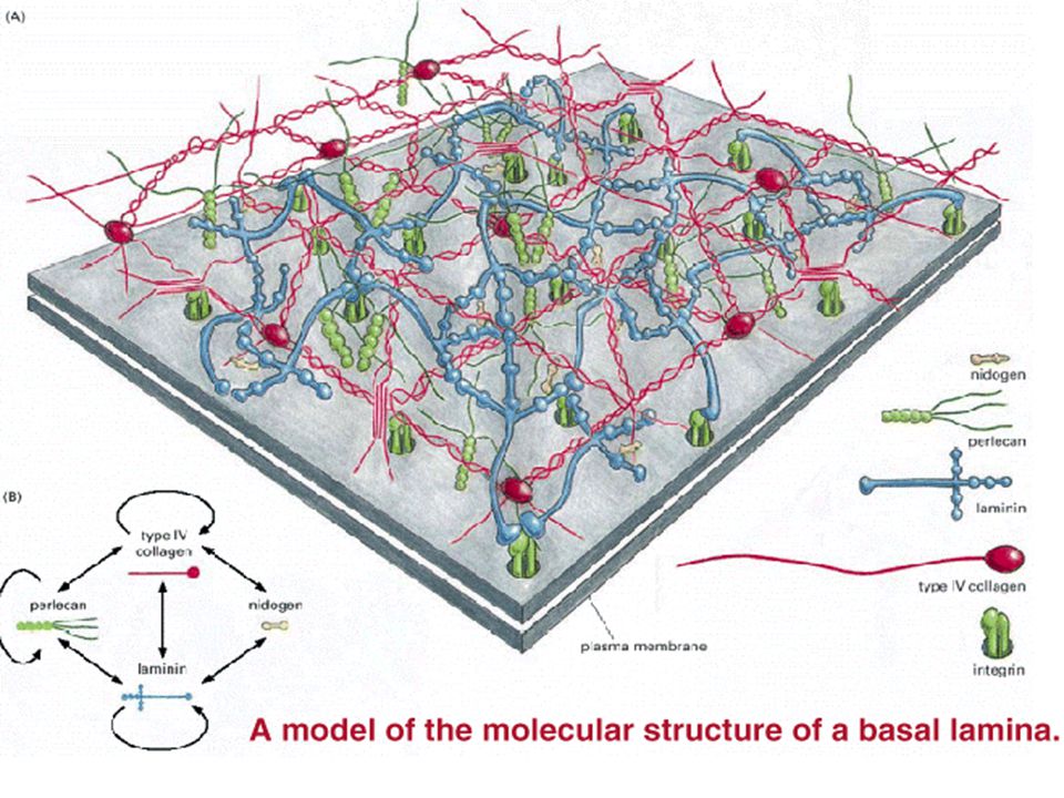
LA MATRICE EXTRACELLULAIRE

38
Coalignment of extracellular fibronectin filaments and intracellular actin filament bundles. (A). The fibronectin is visualized in two rat fibroblasts in culture by the binding of rhodamine- coupled anti- fibronectin antibodies. (B). The actin is visualized by the binding of fluorescein- coupled anti- actin antibodies.
. The fibronectin is visualized in two rat fibroblasts in culture by the binding of rhodamine- coupled anti- fibronectin antibodies. (B). The actin is visualized by the binding of fluorescein- coupled anti- actin antibodies.")
41
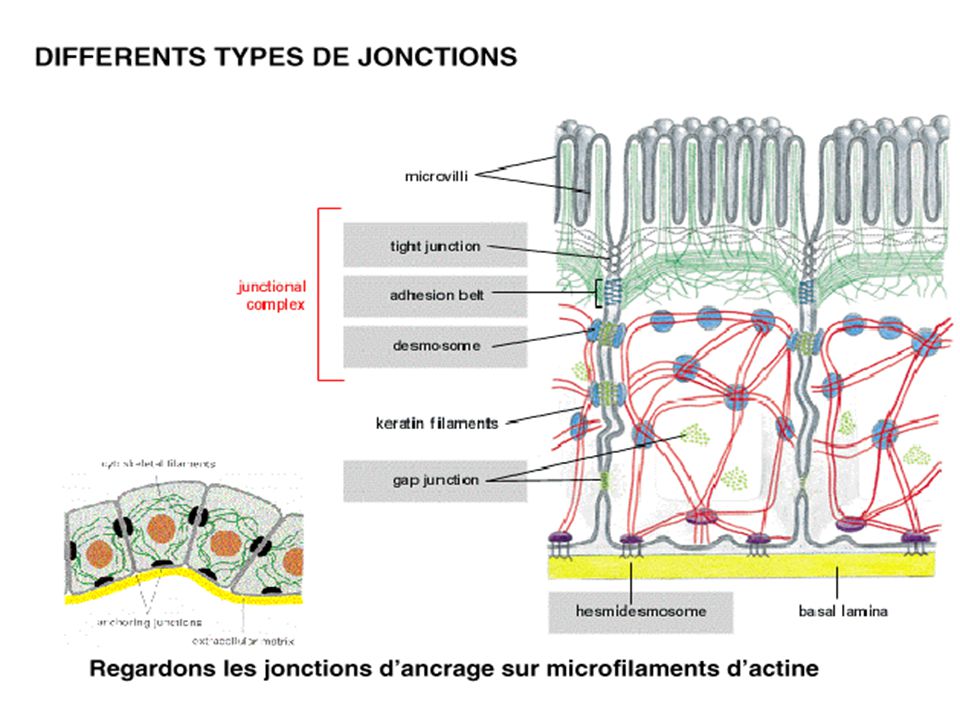
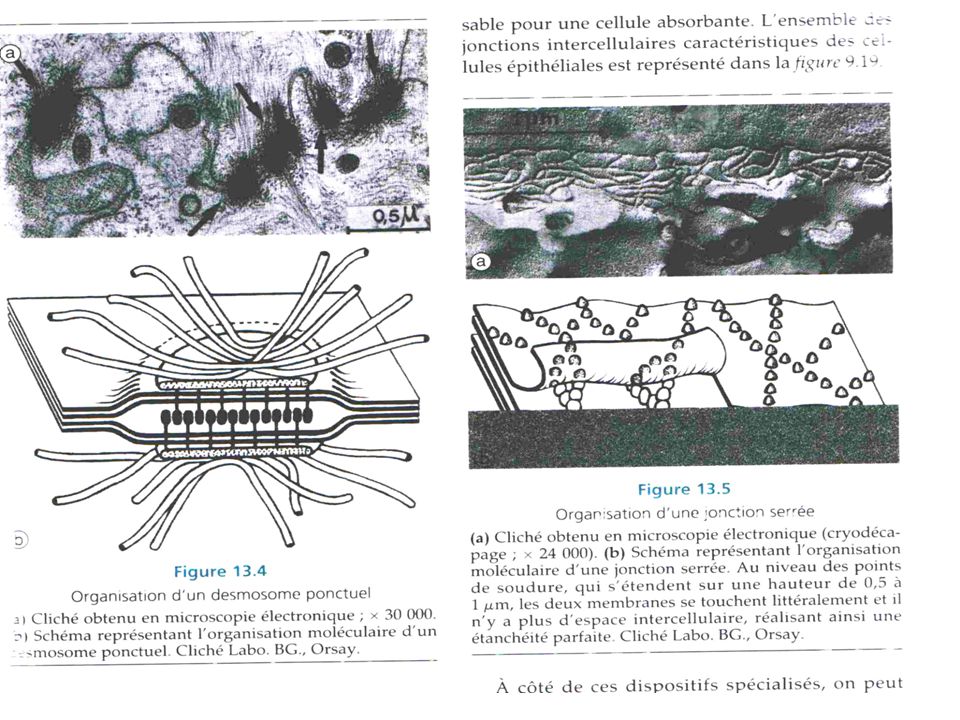
DIFFERENTS TYPES DE JONCTIONS

44
cytoplasmic fracture face of the membrane (the P face) or as
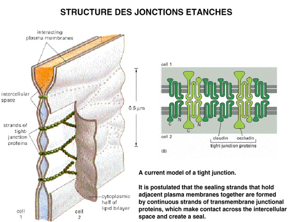
Structure of a tight junction between epithelial cells of the small intestine. The junctions are shown schematically in (A) and in freeze- fracture (B) and conventional (C) electron micrographs. In (B) the tight junction appears as a beltlike band of anastomosing sealing strands that encircle each cell in the sheet. The sealing strands are seen as ridges of intramembrane particles on the cytoplasmic fracture face of the membrane (the P face) or as complementary grooves on the external face of the membrane (the E face). In (C) the junction is seen as a series of focal connections between the outer leaflets of the two interacting plasma membranes, each connection corresponding to a sealing strand in cross- section.
 and in freeze- fracture (B) and conventional (C) electron micrographs. In (B) the tight junction appears as a beltlike band of anastomosing sealing strands that encircle. each cell in the sheet. The sealing strands are seen as ridges of intramembrane particles on the. cytoplasmic fracture face of the membrane (the P face) or as. complementary grooves on the external face of the membrane (the E face). In (C) the junction is. seen as a series of focal connections between the outer leaflets of the two interacting plasma. membranes, each connection corresponding to a sealing strand in cross- section.")
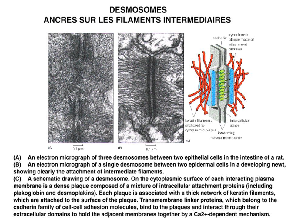
45
Les jonctions septales des invertébrés
A electron micrograph of a septate junction between two epithelial cells in a mollusk. The interacting plasma membranes, seen in cross section, are connected by parallel rows of junctional proteins. The rows, which have a regular periodicity, are seen as dense bars, or septa. ( John Wiley & Sons, Inc .)
")
51
The regulation of gap- junction coupling by a neurotransmitter .
The regulation of gap- junction coupling by a neurotransmitter . (A) A neuron in a rabbit retina was injected with the dye Lucifer yellow, which passes readily through gap junctions and labels other neurons of the same type that are connected to the injected cell by gap junctions. (B) The retina was first treated with the neurotransmitter dopamine, before the neuron was injected with dye. As can be seen, the dopamine treatment greatly decreased the permeability of the gap junctions. ( David Vaney .)
 A neuron in a rabbit retina was injected with the dye Lucifer yellow, which passes. readily through gap junctions and labels other neurons of the same type that are connected to. the injected cell by gap junctions. (B) The retina was first treated with the neurotransmitter dopamine, before the neuron was. injected with dye. As can be seen, the dopamine treatment greatly decreased the. permeability of the gap junctions. ( David Vaney .)")
52
fracture face (P face) of the plasma membrane.
Thin- section (A) and freeze- fracture (B) electron micrographs of a large and a small gap junction between fibroblasts in culture. In (B) each gap junction is seen as a cluster of homogeneous intramembrane particles associated exclusively with the cytoplasmic fracture face (P face) of the plasma membrane. Each intramembrane particle corresponds to a connexon .
 and freeze- fracture (B) electron micrographs of a large and a small. gap junction between fibroblasts in culture. In (B) each gap junction is seen as a cluster. of homogeneous intramembrane particles associated exclusively with the cytoplasmic. fracture face (P face) of the plasma membrane. Each intramembrane particle corresponds to a connexon .")
53
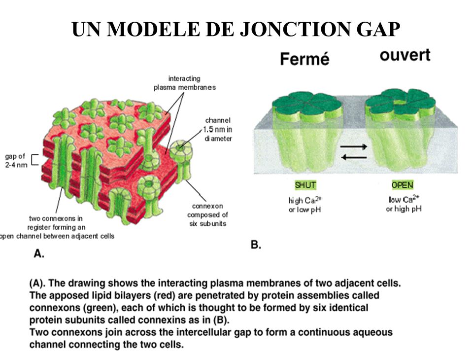
UN MODELE DE JONCTION GAP
Présentations similaires