Télécharger la présentation
La présentation est en train de télécharger. S'il vous plaît, attendez

1
Physiopathologie des infections à P. aeruginosa
DU Antibiothérapie St Louis-Mars 2008 B. Guery EA 2689

3
La virulence de P.aeruginosa
Q U O R M S E N I N G SSTT Pili Flagelle Biofilm Alginate Lectines Mécanismes intriqués

4
Les deux modes de virulences de P. aeruginosa
Infection aiguë Infection chronique Infection invasive caractérisée par la sécrétion de nombreuses toxines (injectisome) Infection inflammatoire associée à la capacité de P. aeruginosa à former une communauté appelée biofilm
 Infection inflammatoire associée à la capacité de P. aeruginosa à former une communauté appelée biofilm.")
5
Les deux modes de virulences de P. aeruginosa
Colonisation initiale Le choix Étape infectieuse Infection aiguë Infection chronique

6
Lung infection model in Pseudomonas aeruginosa
Active TTSS TTSS switched on Toxic effectors TTSS switched off Lung epithelium Polack et al

7
Étape d’adhésion initiale
Reconnaissance de structures glycaniques épithéliales et muciniques via des lectines Colonisation pulmonaire Formation du biofilm

8
Les lectines de P. aeruginosa
(Cioci et al., 2003) LecA (PA-IL) (Loris et al., 2003) LecB (PA-IIL) D-galactose L-fucose L’expression de LecA et LecB est régulée par le système du quorum sensing et par le facteur RpoS
 LecA (PA-IL) (Loris et al., 2003) LecB (PA-IIL) D-galactose. L-fucose. L’expression de LecA et LecB est régulée par le système du quorum sensing et par le facteur RpoS.")
9
Implication des lectines LecA et LecB dans la pathogénie de P
Implication des lectines LecA et LecB dans la pathogénie de P. aeruginosa Etudes in vitro LecA: activité cytotoxique (Bajolet-Laudinat et al., 1994; Wu et al., 2003) LecA et LecB: formation du biofilm (Tielker, al., 2005 ; Diggle et al., 2006) LecA et LecB: inhibition des battements ciliaires (Mewe et al., 2005) Etudes in vivo
 LecA et LecB: formation du biofilm (Tielker, al., 2005 ; Diggle et al., 2006) LecA et LecB: inhibition des battements ciliaires (Mewe et al., 2005) Etudes in vivo.")
10
Chemani et al, Soumis

11
Chemani et al, Soumis

12
Chemani et al, Soumis

13
Chemani et al, Soumis

14
Chemani et al, Soumis

15
Chemani et al, Soumis

16
Capacité à former un biofilm
Quantification of biofilm formation by the PAK, lecA mutant, lecB mutant, complemented lecB mutant (lecB), and PAK-NP strains in a crystal violet microtiter plate assay after 24 h. Absorbance was measured at 600 nm. The means standard deviation (error bars) were determined from at least three independent experiments. The difference between the parent strain and lecB mutant was statistically significant (P 0.05). Pseudomonas aeruginosa expresses two lectins which are implicated in adhesion and biofilm formation. In this study, we demonstrate that P. aeruginosa LecB is involved in pilus biogenesis and proteolytic activity. Moreover, neither lectin was involved in adhesion to human tracheobronchial mucin. We infer that some of the ascribed functions are secondary effects on other systems rather than effects of the lectins themselves. Sonawane et al, Inf Imm 2006
, and PAK-NP strains in a crystal violet microtiter plate assay after 24 h. Absorbance was measured at 600 nm. The means standard deviation (error bars) were determined from at least three independent experiments. The difference between the parent strain and lecB mutant was statistically significant (P 0.05). Pseudomonas aeruginosa expresses two lectins which are implicated in adhesion and biofilm formation. In. this study, we demonstrate that P. aeruginosa LecB is involved in pilus biogenesis and proteolytic activity. Moreover, neither lectin was involved in adhesion to human tracheobronchial mucin. We infer that some of the. ascribed functions are secondary effects on other systems rather than effects of the lectins themselves. Sonawane et al, Inf Imm")
17
Facteurs extracellulaires de virulence
Lazdunski Ann Fr Anesth Réanim 2003,22,523

19
Elastase et fonction pulmonaire
Dose Time T° The alveolar epithelium is lined by surfactant, a lipoprotein complex that both reduces surface tension and mediates several innate immune functions including bacterial aggregation, alteration of alveolar macrophage function, and regulation of bacterial clearance. Surfactant protein-D (SP-D) participates in several of these immune functions, and specifically it enhances the clearance of the pulmonary pathogen Pseudomonas aeruginosa, a common cause of morbidity and mortality in cystic fibrosis (CF) patients. P. aeruginosa secretes a variety of virulence factors including elastase, a zinc-metalloprotease, which degrades both SP-A and SP-D. Here we show that SP-D is cleaved by elastase to produce a stable 35-kDa fragment in a time-, temperature-, and dose-dependent manner. Degradation is inhibited by divalent metal cations, a metal chelator, and the elastase inhibitor, phosphoramidon. Sequencing the SP-D degradation products localized the major cleavage sites to the C-terminal lectin domain. The SP-D fragment fails to bind or aggregate bacteria that are aggregated by intact SP-D. SP-D fragment is observed when normal rat bronchoalveolar lavage (BAL) is treated with Pseudomonas aeruginosa elastase, and SP-D fragments are present in the BAL of CF lung allograft patients. These data show that degradation of SP-D occurs in the BAL environment and that degradation eliminates many normal immune functions of SP-D. The SP-D fragment fails to bind or aggregate bacteria that are aggregated by intact SP-D Alcorn et al, J Biol Chem 2004
 participates in several of these immune functions, and specifically it enhances the clearance of the pulmonary pathogen Pseudomonas aeruginosa, a common cause of morbidity and mortality in cystic fibrosis (CF) patients. P. aeruginosa secretes a variety of virulence factors including elastase, a zinc-metalloprotease, which degrades both SP-A and SP-D. Here we show that SP-D is cleaved by elastase to produce a stable 35-kDa fragment in a time-, temperature-, and dose-dependent manner. Degradation is inhibited by divalent metal cations, a metal chelator, and the elastase inhibitor, phosphoramidon. Sequencing the SP-D degradation products localized the major cleavage sites to the C-terminal lectin domain. The SP-D fragment fails to bind or aggregate bacteria that are aggregated by intact SP-D. SP-D fragment is observed when normal rat bronchoalveolar lavage (BAL) is treated with Pseudomonas aeruginosa elastase, and SP-D fragments are present in the BAL of CF lung allograft patients. These data show that degradation of SP-D occurs in the BAL environment and that degradation eliminates many normal immune functions of SP-D. The SP-D fragment fails to bind. or aggregate bacteria that are. aggregated by intact SP-D. Alcorn et al, J Biol Chem")
20
TTSS: a needle Kubori et al. Science 1998,280,602

21
E. Kipnis et al., Med Mal Infect

23
Exo T ExoT inhibe l’internalisation de PA103 par les cellules épithéliales polarisées et les macrophages Action liée à une structure en doigt d’arginine, mutation: Diminue l’activité anti-internalisation Altération de la capacité à altérer le squelette actino-myosique Garrity-Ryan et al, Inf Imm 2000

24
Exo S et lésion alvéolaire
Percentage of alveolar tracer Lung (% inst) Blood (% inst) Ctr /-2 3+/-2 PAK (A+,S+) 80+/ /-4 PAKexsA (A+,S-) 91+/-2 6+/-1 PA103 (A+,S+) 56+/ /-7 PA103exsA (A-,S-) 89+/-3 5+/-3 (A+,S+) 54+/ /-12 Alv epith injury depends on exoenz S (Kudoh et al, Am J Physiol 1994)
 Blood (% inst) Ctr 93+/-2 3+/-2. PAK (A+,S+) 80+/-4 16+/-4. PAKexsA (A+,S-) 91+/-2 6+/-1. PA103 (A+,S+) 56+/-2 21+/-7. PA103exsA (A-,S-) 89+/-3 5+/-3. (A+,S+) 54+/ /-12. Alv epith injury depends on exoenz S. (Kudoh et al, Am J Physiol 1994)")
25
Mutation sur l’activité ADRBT Shaver & Hauser, Inf Imm 2004
Pseudomonas aeruginosa uses a type III secretion system to promote development of severe disease, particularly in patients with impaired immune defenses. While the biochemical and enzymatic functions of ExoU, ExoS, and ExoT, three effector proteins secreted by this system, are well defined, the relative roles of each protein in the pathogenesis of acute infections is not clearly understood. Since ExoU and ExoS are usually not secreted by the same strain, it has been difficult to directly compare the effects of these proteins during infection. In the work described here, several isogenic mutants of a bacterial strain that naturally secretes ExoU, ExoS, and ExoT were generated to carefully evaluate the relative contribution of each effector protein to pathogenesis in a mouse model of acute pneumonia. Measurements of mortality, bacterial persistence in the lung, and dissemination indicated that secretion of ExoU had the greatest impact on virulence while secretion of ExoS had an intermediate effect and ExoT had a minor effect. It is of note that these results conclusively show for the first time that ExoS is a virulence factor. Infection with isogenic mutants secreting wild-type ExoS, ExoS defective in GTPase-activating protein (GAP) activity, or ExoS defective in ADP-ribosyltransferase activity demonstrated that the virulence of ExoS was largely dependent on its ADP-ribosyltransferase activity. The GAP activity of this protein had only a minor effect in vivo. The relative virulence associated with each of these type III effector proteins may have important prognostic implications for patients infected with P. aeruginosa. Mutation sur l’activité ADRBT Shaver & Hauser, Inf Imm 2004
 activity, or ExoS defective in ADP-ribosyltransferase activity demonstrated that the virulence of ExoS was largely dependent on its ADP-ribosyltransferase activity. The GAP activity of this protein had only a minor effect in vivo. The relative virulence associated with each of these type III effector proteins may have important prognostic implications for patients infected with P. aeruginosa. Mutation sur l’activité ADRBT. Shaver & Hauser, Inf Imm")
26
CHO cell Phillips et al, JBC 2003
Cytotoxicity of purified ExoU. a, buffer alone, BSA (50 µg), ExoU (50 µg), or papain-digested ExoU (50 µg) was syringe-loaded into CHO cells, and cytotoxicity was measured by trypan blue exclusion (percent of cell death, gray) or LDH activity (white, 100 x A490), with LDH activity from buffer alone subtracted. Error bars indicate the standard deviation of triplicate experiments for this and the following figures. b, cytotoxic dose-response in CHO cells, as measured by LDH activity. ExoU was quantified using an experimentally determined absorption extinction coefficient ( 280 = 31,200 M–1 cm–1) and CHO cells by counting trypan blue-excluding cells. As shown in c, ExoU(S142A)-GFP localizes to the plasma membrane in punctate fashion. A fluorescence microscopy image of CHO cells syringe-loaded with ExoU(S142A)-GFP or GFP is shown. Cell nuclei are visualized by Hoechst staining (blue), and GFP fluorescence is seen in green. For ExoU(S142A)-GFP, representative focal planes are shown to demonstrate circumferential plasma membrane (Focal Plane 1) and punctate localization (Focal Plane 2). CHO cell Phillips et al, JBC 2003
, ExoU (50 µg), or papain-digested ExoU (50 µg) was syringe-loaded into CHO cells, and cytotoxicity was measured by trypan blue exclusion (percent of cell death, gray) or LDH activity (white, 100 x A490), with LDH activity from buffer alone subtracted. Error bars indicate the standard deviation of triplicate experiments for this and the following figures. b, cytotoxic dose-response in CHO cells, as measured by LDH activity. ExoU was quantified using an experimentally determined absorption extinction coefficient ( 280 = 31,200 M–1 cm–1) and CHO cells by counting trypan blue-excluding cells. As shown in c, ExoU(S142A)-GFP localizes to the plasma membrane in punctate fashion. A fluorescence microscopy image of CHO cells syringe-loaded with ExoU(S142A)-GFP or GFP is shown. Cell nuclei are visualized by Hoechst staining (blue), and GFP fluorescence is seen in green. For ExoU(S142A)-GFP, representative focal planes are shown to demonstrate circumferential plasma membrane (Focal Plane 1) and punctate localization (Focal Plane 2). CHO cell. Phillips et al, JBC")
27
Effects of PLA2 inhibitors (a and b) or non-PLA2 inhibitors (c) in syringe-loading experiments or infection assays. As shown in a and c, inhibitors were syringe-loaded with ExoU (0.675 µM) into CHO cells and included in culture medium afterward. Inhibitors were used at the following concentrations: MAFP, 67.5 µM; ATK, 67.5 µM; PTK, 67.5 µM; LY311727, 250 µM; pyrrophenone, 1 µM; bromoenol lactone (BEL), 67.5 µM; phenylmethylsulfonyl fluoride (PMSF), 1.0 mM; genistein, 200 µM; wortmannin, 100 nM; PD98059, 50 µM; nordihydroguaiaretic acid (NDGA), 100 µM; and aspirin, 100 µM. b, MAFP inhibition of cytotoxicity in CHO cells infected with P. aeruginosa PA103, as assessed by LDH activity. Background LDH activity from medium alone is shown and was not subtracted from sample values. Phillips et al, JBC 2003

28
Lung epithelial injury (%131I-Alb) Cytotoxicity (%)
Mice instilled with 1.10^7 CFU/ml PA103 : parental strain dPcrV : souche mutée sur PcrV dPcrV/pUCPpcrV : souche mutée sur Pcrv complémentée par un plasmide exprimant PcrV dPcrV/pUCP : souche mutée su PcrV complémentée par un plasmide controle (Sawa et al, Nature Med 1999)
")
29
MAP Cardiac Output (mmHg) (% baseline) Time (hours) Time (hours)
Airspace instillation of PA103 dUT : mutant présentant un défect sur le syst de sécretion de type III Time (hours) Time (hours) (Kurahashi et al, J Clin Invest 1999)
 Time (hours) (Kurahashi et al, J Clin Invest 1999)")
30
MAP Cardiac Output (mmHg) (% baseline) Time (hours) Time (hours)
(Kurahashi et al, J Clin Invest 1999)
")
31
125I-alb in blood (%) 125I-alb in blood (%) Time (hours) Time (hours)
Concl : Injury to alv epith allows the release of proinflamm mediators into the circulation that are primaraly responsible for septic shock. Importance of the compartmentalization Time (hours) Time (hours) (Kurahashi et al, J Clin Invest 1999)
 Time (hours) (Kurahashi et al, J Clin Invest 1999)")
32
Surmortalité et SST III
Infection aiguë Infection chronique SSTT [+] 89% 41% SSTT [+] SSTT [-] Mortalité 21% 3% The ability of Pseudomonas aeruginosa to secrete specific toxins using the type III-mediated pathway has been reported. To determine the association of this phenotype with human illness, immunoblot analysis was used to detect expression of type III secretory proteins in P. aeruginosa isolates from respiratory tract or blood cultures of 108 consecutive patients. Relative risk of mortality was 6-fold greater with expression of the type III secretory proteins ExoS, ExoT, ExoU, or PcrV. Phenotype was independently correlated with toxicity in cellular and murine models. Prevalence of this phenotype was significantly higher in acutely infected patients than in chronically infected patients with cystic fibrosis. These results suggest that the type III protein secretion system is integral to increased P. aeruginosa virulence. A positive phenotype is a predictor of poor clinical outcome. In the future, such analyses may help distinguish potentially lethal infection from colonization and help determine appropriate therapy for critically ill patients. RR décès PcrV seule 7,4 PcrV + toxine(s) 8,7 (Roy Burman et al, J Infect Dis )
 8,7. (Roy Burman et al, J Infect Dis )")
33
Pneumonie à P.aeruginosa
35 patients ventilés Pneumonie à P.aeruginosa Production de Protéines Issues du système de sécrétion de type III + 27/35 - 8/35 22 Sévère 5 Modérées 3 Sévères 5 Modérées (81%) (19%) (38%) (62%) ExoU : 10/35 (29%) associée à 90% de formes sévères (Hauser et al, Crit Care Med 2002)
 (19%) (38%) (62%) ExoU : 10/35 (29%) associée à 90% de formes sévères. (Hauser et al, Crit Care Med 2002)")
34
Macnab Annu Rev Microbiol 2003,57:77
Flagelle et SSTT Macnab Annu Rev Microbiol 2003,57:77

35
La flagelline (FliC)
")
36
In this study, we tested the contribution of flagellar motility, flagellin structure, and its glycosylation in Pseudomonas aeruginosa using genetically defined flagellar mutants. All mutants and their parent strains were tested in a burned-mouse model of infection. Motility and glycosylation of the flagellum appear to be important determinants of flagellar-mediated virulence in this model. This is the first report where genetically defined flagellar variants of P. aeruginosa were tested in the burned-mouse model of infection. Arora et al, Inf Imm 2005

37
PILI

38
Retraction des pilis

39
Tang H et al, Infect Immun 1995
infant BALB/cByJ The role of piliation in the development and course of acute pulmonary infection was examined using infant BALB/cByJ mice inoculated by intranasal instillation of isogenic Pil+ and Pil- mutants of Pseudomonas aeruginosa PA1244, PAK, and PAO1. The piliated strains caused more cases of pneumonia, bacteremia, and mortality than the nonpiliated strains (chi-square analysis, alpha = 0.001). The piliated strains were more often associated with severe diffuse pneumonias, while the nonpiliated organisms resulted in less severe, focal pneumonias, although these differences did not achieve statistical significance. Purified pilin protein used to inoculate the mice resulted in local inflammatory changes. The nonpiliated strain PA1244-NP was as virulent as the piliated strain PAO1, suggesting that expression of other virulence factors are also important in the development of acute pneumonia. This infant mouse model of pulmonary infection appears to be a useful system for the analysis of P. aeruginosa virulence factors involved in the pathogenesis of pneumonia. Pourquoi les pilines? Interactions entre récepteurs épithéliaux et pilines. Tx de ce récepteur augmenté +++ chez muco Intranasal instillation mice Balb/c, 3 souches Isogenic mutant Pil+ or Pil- Pil+ causes more mortality, bacteremia and pneumonia and more diffuse pneumonia NO STATISTICAL DIFFERENCE sauf PAO1/pneumonie et PA1244/mortalité Inoculation de piline purifiée caused local inflammation Différences entre les 3 souches +++ Gain + marqué pour PAO1 Conclusion: other virulence factors are important Perspectives: prophylaxie dans la muco, stade précoce: colonisation? Tang H et al, Infect Immun 1995
. The piliated strains were more often associated with severe diffuse pneumonias, while the nonpiliated organisms resulted in less severe, focal pneumonias, although these differences did not achieve statistical significance. Purified pilin protein used to inoculate the mice resulted in local inflammatory changes. The nonpiliated strain PA1244-NP was as virulent as the piliated strain PAO1, suggesting that expression of other virulence factors are also important in the development of acute pneumonia. This infant mouse model of pulmonary infection appears to be a useful system for the analysis of P. aeruginosa virulence factors involved in the pathogenesis of pneumonia. Pourquoi les pilines Interactions entre récepteurs épithéliaux et pilines. Tx de ce récepteur augmenté +++ chez muco. Intranasal instillation mice Balb/c, 3 souches. Isogenic mutant Pil+ or Pil- Pil+ causes more mortality, bacteremia and pneumonia and more diffuse pneumonia. NO STATISTICAL DIFFERENCE sauf PAO1/pneumonie et PA1244/mortalité. Inoculation de piline purifiée caused local inflammation. Différences entre les 3 souches +++ Gain + marqué pour PAO1. Conclusion: other virulence factors are important. Perspectives: prophylaxie dans la muco, stade précoce: colonisation Tang H et al, Infect Immun")
40
alginate

41
Alginate mAb humanisé Strain MAb Survie Ctr Survie mAb N13 F429g1 0/6
Non mucoide Strain MAb Survie Ctr Survie mAb N13 F429g1 0/6 6/6 F428g1 0/10 8/10 PAO1 4/6 PA14 5/5 PAO1 ExoU+ 0/5 B312 Two fully human mAbs specific for epitopes dependent on intact carboxylate groups on the C6 carbon of the mannuronic acid components of Pseudomonas aeruginosa alginate were found to promote phagocytic killing of both mucoid and nonmucoid strains as well as protection against both types of strains in a mouse model of acute pneumonia. The specificity of the mAbs for alginate was determined by ELISA and killing assays. Some strains of P. aeruginosa did not make detectable alginate in vitro, but in vivo protection against lethal pneumonia was obtained and shown to be due to rapid induction of expression of alginate in the murine lung. No protection against strains genetically unable to make alginate was achieved. These mAbs have potential to be passive therapeutic reagents for all strains of P. aeruginosa and the results document that alginate is a target for the proper type of protective Ab even when expressed at low levels on phenotypically nonmucoid strains. Pier et al, J Immunol 2004

42
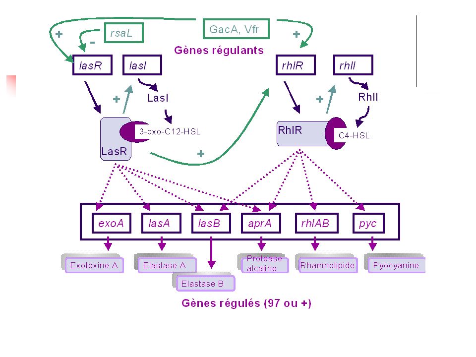
Quorum Sensing

43
Faible Concentration en AHL Forte Concentration en AHL
luxR luxI luxR luxI LuxR LuxI LuxR LuxI AHL AHL AHL AHL AHL AHL AHL AHL Le gène luxI code pour une enzyme qui intervient dans la synthèse d’un auto-inducteur de la famille des AHL. Ces molécules diffusent librement au travers des membranes bactériennes et permettent la communication entre les bactéries. Lorsque leur concentration atteint un seuil critique, elles se fixent sur les protéines LuxR codées par le gène luxR. Le complexe LuxR/AHL devient activateur transcriptionnel de gènes impliqués dans la synthèse de molécules luminescentes et de luxI qui synthétise alors plus d’autoinducteur (Figure 1). Transcription & translation LuxR Operon lux Operon lux AHL RNA polymérase RNA polymérase Luminescence
. Transcription & translation. LuxR. Operon lux. Operon lux. AHL. RNA polymérase. RNA polymérase. Luminescence.")
44
Vfr VqsR QscR RsaL lasR lasI RsaL LasR LasI RhlR RhlI RhlR RhlI PqsR
Activation Inhibition Vfr VqsR RsaL lasR lasI RsaL QscR LasR LasI C12 C12 PQS RhlR RhlI pqsH RhlR RhlI pqsR PqsR C4 C4 pqsA pqsB pqsC pqsD pqsE

46
* * * 109 CFU/souris H18 Mort Pneumonie Bactériémie
FIG. 1. Contribution of P. aeruginosa AHL synthase genes lasI and rhl in the pathogenesis of pneumonia, bacteremia, and mortality in neonatal mice. The percentage of the total number of mice that developed pneumonia (dotted bars), bacteremia (white bars), or died (black bars) by 18 h following intranasal inoculation with the P. aeruginosa strains indicated is shown: PAO1, n = 34; PAO-JP1 n = 24; PDO100, n = 23; PAO-JP2, n = 21; and PAO-JP2(pJPP42), n = 21. P values are indicated in the text. Pearson et al , Inf Imm 2000
, bacteremia (white bars), or died (black bars) by 18 h following intranasal inoculation with the P. aeruginosa strains indicated is shown: PAO1, n = 34; PAO-JP1 n = 24; PDO100, n = 23; PAO-JP2, n = 21; and PAO-JP2(pJPP42), n = 21. P values are indicated in the text. Pearson et al , Inf Imm")
47
Modèle de souris brûlée s/c : Injection 102 CFU
Sur un modèle de souris brulé infecté par P. a, rumbaugh a montré que la virulence chez les animaux ayant reçu une souche mutée sur lasI et rhlI est moindre que chez les souris ayant reçu la souche PAO1 : Mortalité Dissémination bactérienne au niveau du foie et de la rate Rumbaugh et al, Inf Imm 1999

48
Etude Pyopneumagen H4 5 souris/souche (280 souris)
+ cytotoxicité sur A549 5 souris/souche (280 souris) Instillation intratrachéale de bactérie I-Alb Souris BALBc 125 I-Alb Œdème pulmonaire = mouillé/sec H4 125 I-Alb Trouble de perméabilité = 125 I-Alb plasmatique + Hémocultures LeBerre et al
 Instillation intratrachéale de bactérie I-Alb. Souris BALBc. 125 I-Alb. Œdème pulmonaire. = mouillé/sec. H I-Alb. Trouble de perméabilité. = 125 I-Alb plasmatique. + Hémocultures. LeBerre et al.")
49
Le Berre et al, CMI 2008

50
* * * 58 isolats non clonaux LeBerre et al

51
RESULTATS % I125 W/D hémoc+ 50% Ni UST 4,3 4,27
ExoST 6,6 * 4,44 OR= 2,19 ExoU 12,6 * 5,22 * OR= 9 * * p< 0,05 versus ni UST LeBerre et al

52
Communication inter espèce
CFU % consolidation Quantitative pathology and bacteriology of the rat lungs 7 days after infection using the agar beads model. A. The rat lungs co-infected with P. aeruginosa and OF strain CF004 (group 2) showed significantly more consolidation than that infected with P. aeruginosa alone (group 1) or OF alone (group 3) (P < , unpaired t-test). Consolidation indicates accumulation of pulmonary oedema fluid and/or infiltration of inflammatory cells. More consolidation denotes more severe lung damage. B. P. aeruginosa (solid bars) and CF004 (grey bars) bacterial loads (cfu) in the lungs of the three groups. The change in gene expression patterns in response to host environments is a prerequisite for bacterial infection. Bacterial diseases often occur as an outcome of the complex interactions between pathogens and the host. The indigenous, usually non-pathogenic microflora is a ubiquitous constituent of the host. In order to understand the interactions between pathogens and the resident microflora and how they affect the gene expression patterns of the pathogens and contribute to bacterial diseases, the interactions between pathogenic Pseudomonas aeruginosa and avirulent oropharyngeal flora (OF) strains isolated from sputum samples of cystic fibrosis (CF) patients were investigated. Animal experiments using a rat lung infection model indicate that the presence of OF bacteria enhanced lung damage caused by P. aeruginosa. Genome-wide transcriptional analysis with a lux reporter-based promoter library demonstrated that approximately 4% of genes in the genome responded to the presence of OF strains using an in vitro system. Characterization of a subset of the regulated genes indicates that they fall into seven functional classes, and large portions of the upregulated genes are genes important for P. aeruginosa pathogenesis. Autoinducer-2 (AI-2)-mediated quorum sensing, a proposed interspecies signalling system, accounted for some, but not all, of the gene regulation. A substantial amount of AI-2 was detected directly in sputum samples from CF patients and in cultures of most non-pseudomonad bacteria isolated from the sputa. Transcriptional profiling of a set of defined P. aeruginosa virulence factor promoters revealed that OF and exogenous AI-2 could upregulate overlapping subsets of these genes. These results suggest important contributions of the host microflora to P. aeruginosa infection by modulating gene expression via interspecies communications. 4% des gènes de Pseudomonas répondent à OF Rôle de AI2 Duan et al, Molec Microb 2003
 showed significantly more consolidation than that infected. with P. aeruginosa alone (group 1) or OF alone (group 3) (P < , unpaired t-test). Consolidation indicates accumulation of pulmonary. oedema fluid and/or infiltration of inflammatory cells. More consolidation denotes more severe lung damage. B. P. aeruginosa (solid bars) and CF004 (grey bars) bacterial loads (cfu) in the lungs of the three groups. The change in gene expression patterns in response to host environments is a prerequisite for bacterial infection. Bacterial diseases often occur as an outcome of the complex interactions between pathogens and the host. The indigenous, usually non-pathogenic microflora is a ubiquitous constituent of the host. In order to understand the interactions between pathogens and the resident microflora and how they affect the gene expression patterns of the pathogens and contribute to bacterial diseases, the interactions between pathogenic Pseudomonas aeruginosa and avirulent oropharyngeal flora (OF) strains isolated from sputum samples of cystic fibrosis (CF) patients were investigated. Animal experiments using a rat lung infection model indicate that the presence of OF bacteria enhanced lung damage caused by P. aeruginosa. Genome-wide transcriptional analysis with a lux reporter-based promoter library demonstrated that approximately 4% of genes in the genome responded to the presence of OF strains using an in vitro system. Characterization of a subset of the regulated genes indicates that they fall into seven functional classes, and large portions of the upregulated genes are genes important for P. aeruginosa pathogenesis. Autoinducer-2 (AI-2)-mediated quorum sensing, a proposed interspecies signalling system, accounted for some, but not all, of the gene regulation. A substantial amount of AI-2 was detected directly in sputum samples from CF patients and in cultures of most non-pseudomonad bacteria isolated from the sputa. Transcriptional profiling of a set of defined P. aeruginosa virulence factor promoters revealed that OF and exogenous AI-2 could upregulate overlapping subsets of these genes. These results suggest important contributions of the host microflora to P. aeruginosa infection by modulating gene expression via interspecies communications. 4% des gènes de Pseudomonas répondent à OF. Rôle de AI2. Duan et al, Molec Microb")
53
Super nertwork

54
Super régulateur CyaB Adenylate cyclase AMPc CyaA
Cofacteur Régulateur transcriptionnel Vfr Vfr membre de la famille des proteines récepteur de l’AMPc Expression de plus de 100 gènes SSTT Pili type IV Exotoxin A via LasR/LasI ….. Smith et al, Inf Imm 2004

55
IFR 14 Xavier Leroy GREPI Grenoble Bertrand Toussaint Olivier Epaulard Benoit Polack Genève Christian Vandelden EA 2689 Karine Faure Rozen Le Berre Maud Pierre Chanez Chemani

56
16h pi To confirm whether PCN is important in lung infection, we compared the levels of virulence of wild-type P. aeruginosa PA14 and PAO1 and those of the PCN-deficient isogenic mutants of PA14, the phzB1 and mvfR mutants, and those of the PCN-deficient isogenic mutants of PAO1, the phzM and phzS mutants (Table 1), by using an acute mouse pneumonia model of infection (18). The phzB1, phzM, and phzS genes encode enzymes involved in the final steps of the PCN biosynthetic cascade (10), whereas mvfR encodes a LysR-like transcription activator that controls the phnAB and pqs genes that synthesize quorum-sensing quinolones to regulate PCN biosynthesis (2, 6). Adult CD-1 mice were infected intranasally with 107 bacteria. Infected lungs were harvested at 16 h postinoculation, sectioned, and stained with hematoxylin and eosin for histological analysis. Lau et al, Inf Imm 2004
, by using an acute mouse pneumonia model of infection (18). The phzB1, phzM, and phzS genes encode enzymes involved in the final steps of the PCN biosynthetic cascade (10), whereas mvfR encodes a LysR-like transcription activator that controls the phnAB and pqs genes that synthesize quorum-sensing quinolones to regulate PCN biosynthesis (2, 6). Adult CD-1 mice were infected intranasally with 107 bacteria. Infected lungs were harvested at 16 h postinoculation, sectioned, and stained with hematoxylin and eosin for histological analysis. Lau et al, Inf Imm")
Présentations similaires


 d ’éradication du SARM en soins de longue durée>")


>")


: données de la littérature Dr S. Alfandari Service de Réanimation et Maladies Infectieuses.>")

>")


