Télécharger la présentation
La présentation est en train de télécharger. S'il vous plaît, attendez

1
BIOLOGIE DU DEVELOPPEMENT
Université d’Orléans Prof J.L. PICHON Année

2
Langue : Français Éditeur : De Boeck
(2004) (8 BU) Format : Broché pages 2e édition ISBN : Langue : Français Éditeur : Dunod ( ) (5 & 6 BU) Format : Broché pages ISBN : Editions non disponibles
 (8 BU) Format : Broché pages 2e édition. ISBN : Langue : Français Éditeur : Dunod. ( ) (5 & 6 BU) Format : Broché pages. ISBN : Editions non disponibles.")
3
Langue : anglais, Éditeur : Sinauer
(2006) Format : relié pages 8e édition ISBN : X Langue : anglais, Éditeur : Oxford University Press (2006) Format : Broché-551 pages 3e édition ISBN :
 Format : relié pages 8e édition. ISBN : X. Langue : anglais, Éditeur : Oxford University Press (2006) Format : Broché-551 pages 3e édition. ISBN :")
4
Communication intercellulaires au cours du développement

5
INTRODUCTION

6
INDUCTION – COMPETENCE TISSU INDUCTEUR TISSU CIBLE

7
Ectoderme antérieur Ectoderme postérieur

12
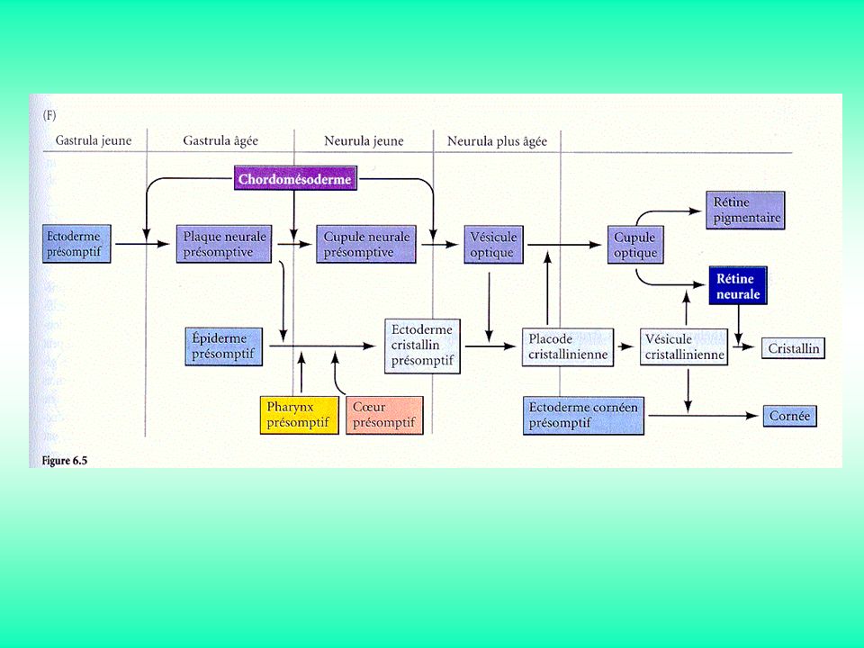
Les signaux inducteurs sont séquentiels et réciproques
Cascades d’induction

15
Interactions de type instructif :
A sur B donne X En absence de A, B ne donne pas X En présence de C, B donne Y Interactions de type permissif : B a tout le nécessaire pour donner X; Il faut seulement un signal de progression

16
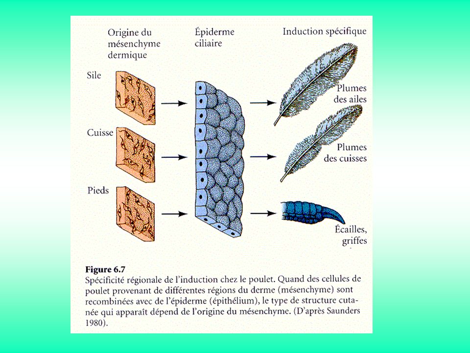
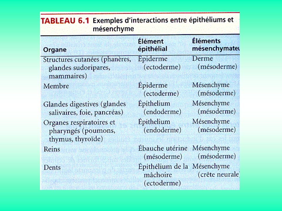
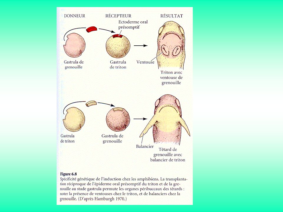
Interactions entre épithélium et mésenchyme

20
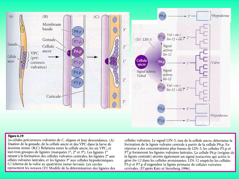
Figure 1 A "hen's tooth" formed by the combination of chick pharygeal (presumptive jaw) ectoderm and mouse molar mesenchyme. (From Kollar and Fisher, 1980; courtesy of E. J. Kollar.)
.")
21
Facteurs Paracrines & Juxtacrines

23
Famille du FGF

25
(Sprouty) (Similar expression to Fgfs) (MAP-kinase-phosphatase 3) (Isthmic organizer) Brain Res Rev (2005)
 (Similar expression to Fgfs) (MAP-kinase-phosphatase 3) (Isthmic organizer) Brain Res Rev (2005)")
27
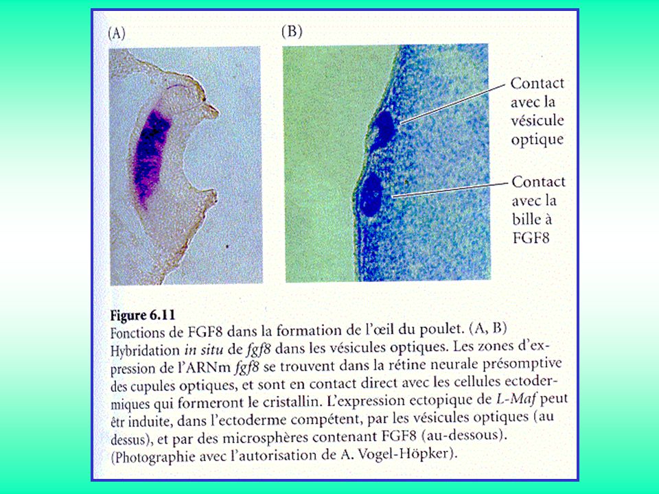
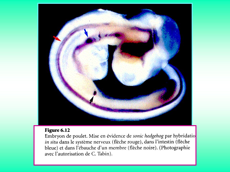
Famille Hedgehog

29
(wingless & integrated)
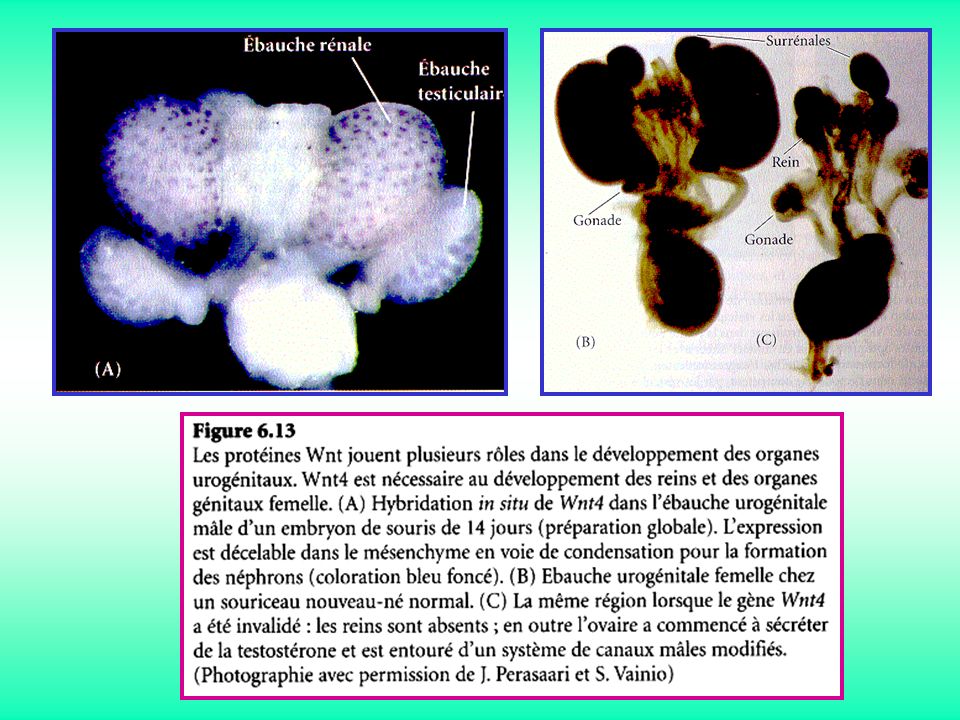
Famille WNT (wingless & integrated)
")
31
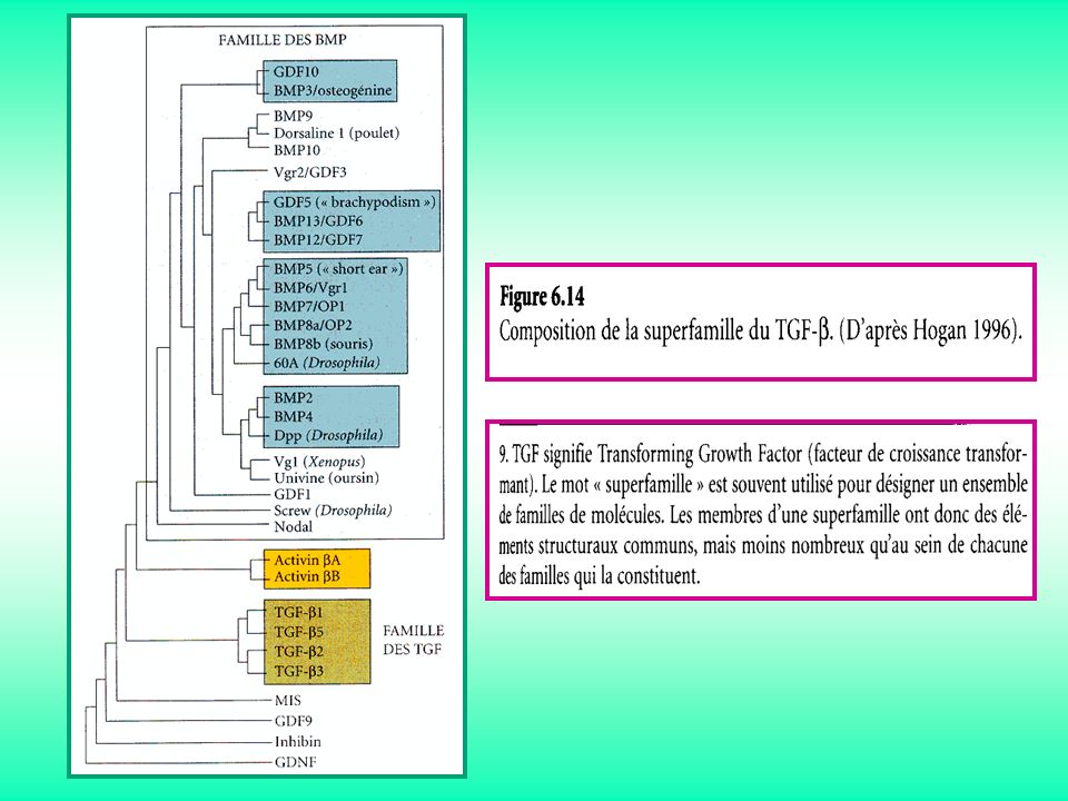
Superfamille TGF b

34
Récepteurs et voies de transmission des signaux

35
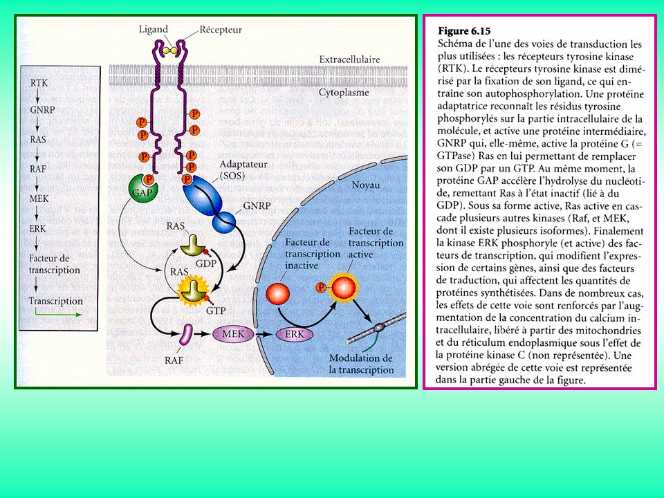
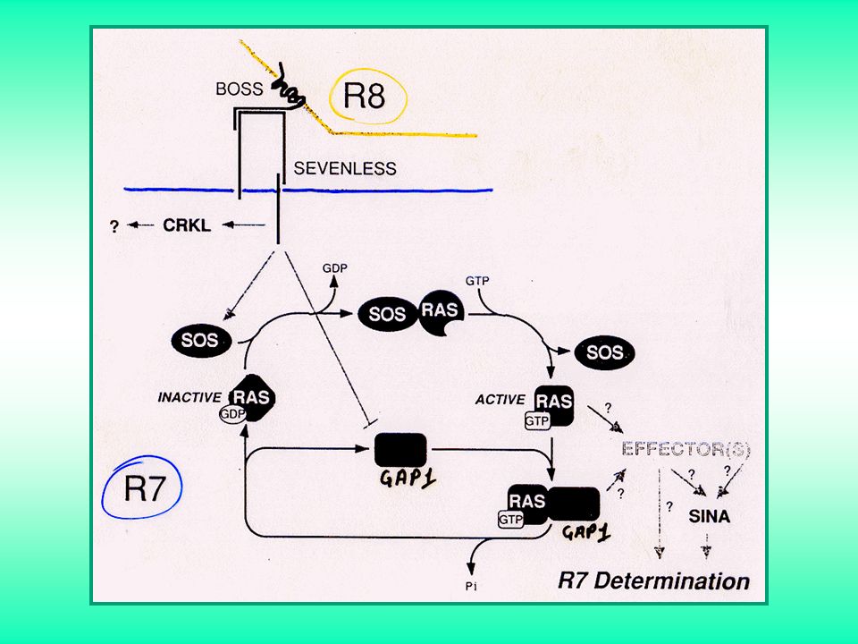
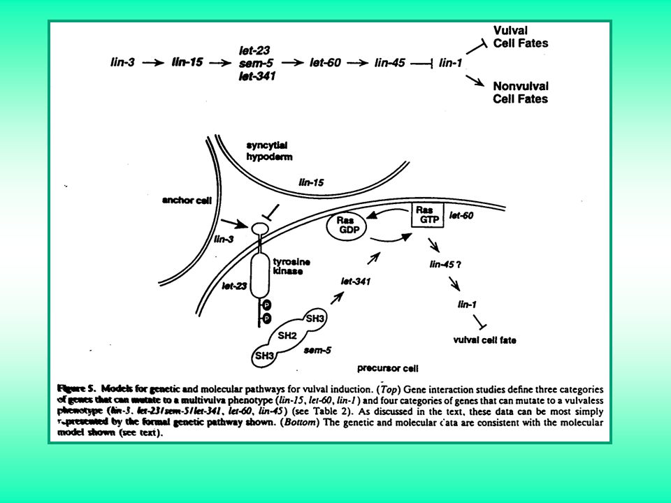
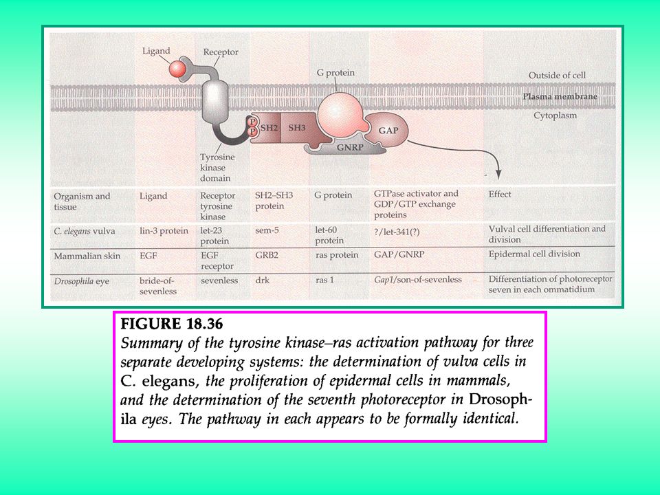
Voie RTK

47
(Sprouty) (Similar expression to Fgfs) (MAP-kinase-phosphatase 3) (Isthmic organizer) Brain Res Rev (2005)
 (Similar expression to Fgfs) (MAP-kinase-phosphatase 3) (Isthmic organizer) Brain Res Rev (2005)")
48
(Heparan sulfate proteoglycan)
Figure 1 Model of interaction between FGF, its receptor, and the proteoglycan carbohydrate groups. Data support the hypothesis that HSPGs modulate the binding of FGF-2 to FGFR through the formation of a ternary complex in which the glycosaminoglycan chain interactswith FGF-2 via 2-O- and N-sulfate groups while 6-O-sulfate groups are required for its interaction with FGFR. A single molecule of heparin/HS may bind several molecules of FGF-2 suggesting that GAG induces oligomerization of FGF-2. (After Presta, M. 2000).
.")
49
Voie Smad

51
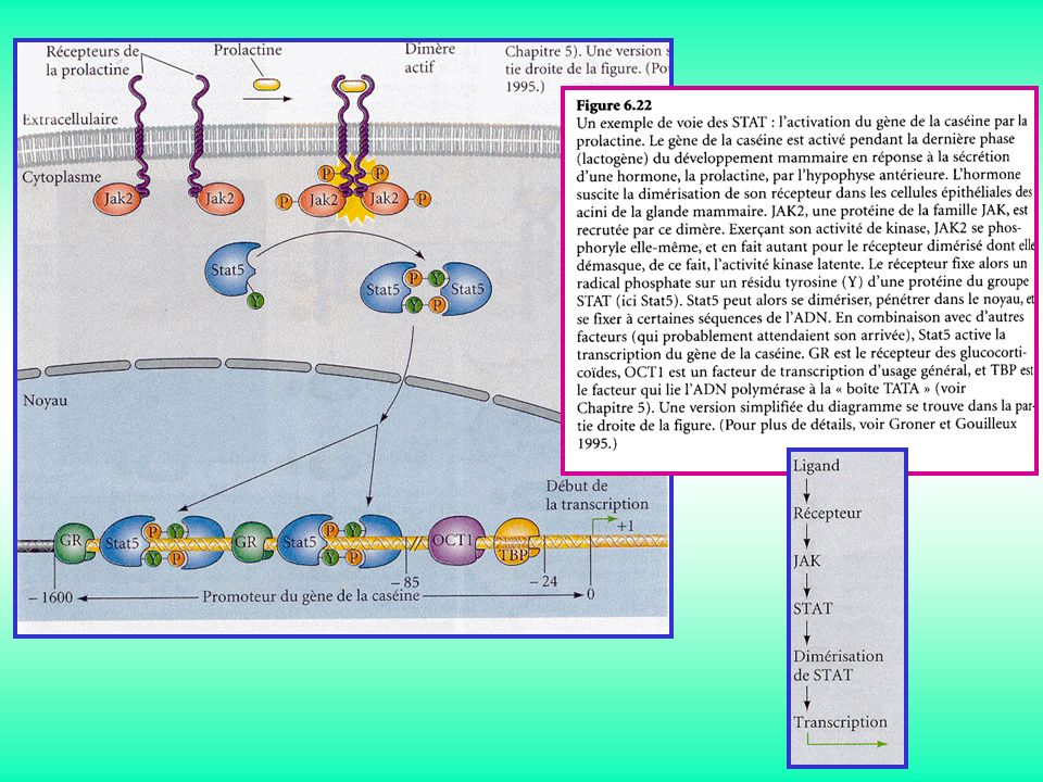
Voie JAK-STAT

54
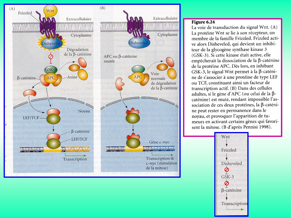
Voie Wnt

56
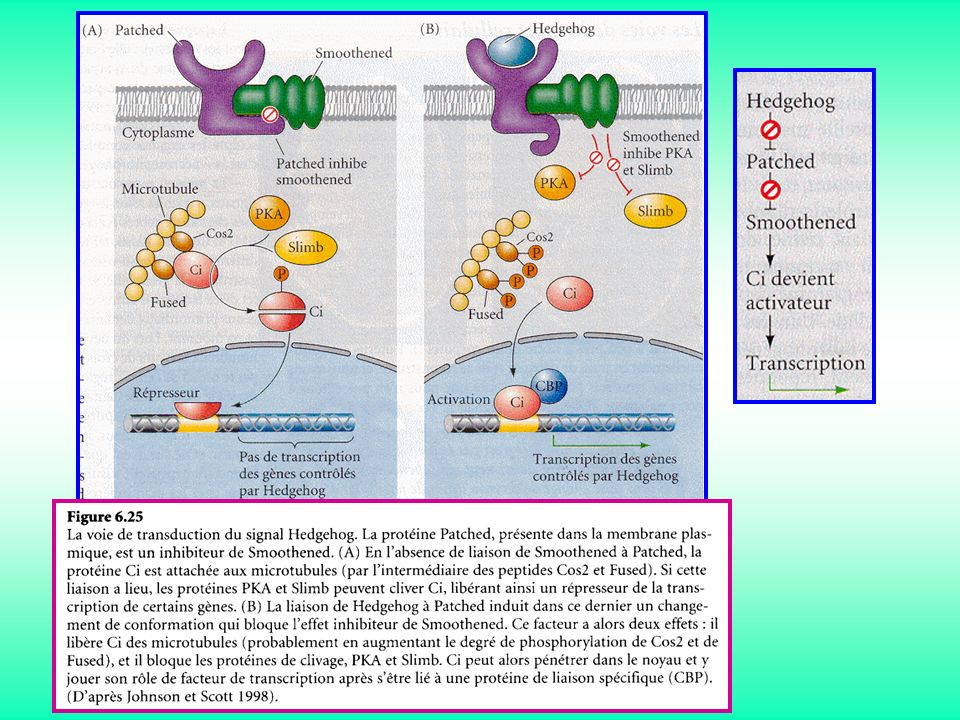
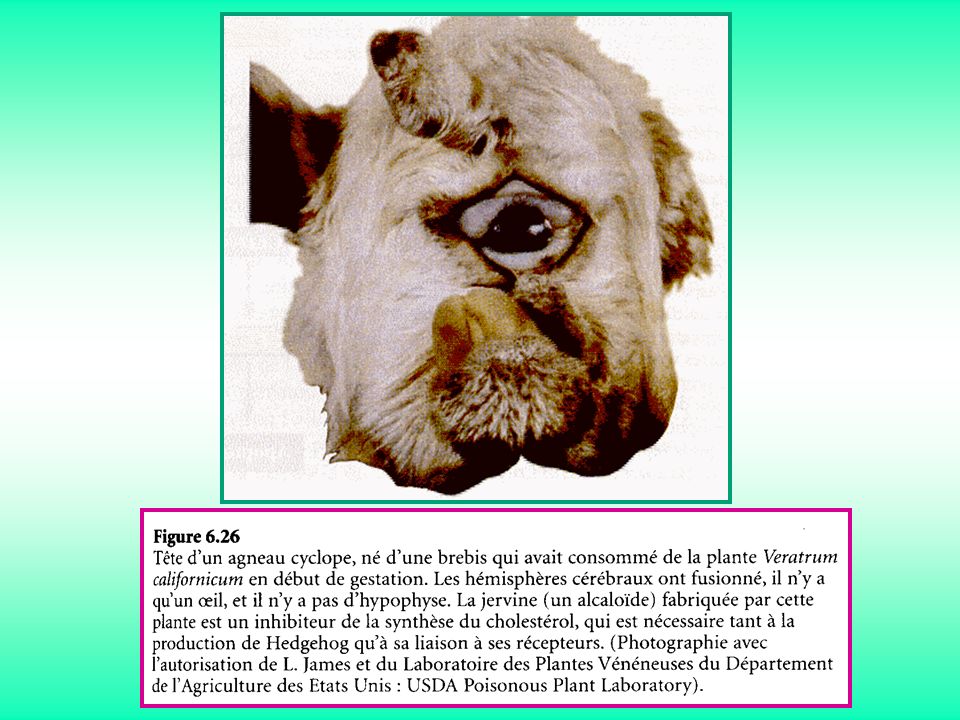
Voie Hedgehog

59
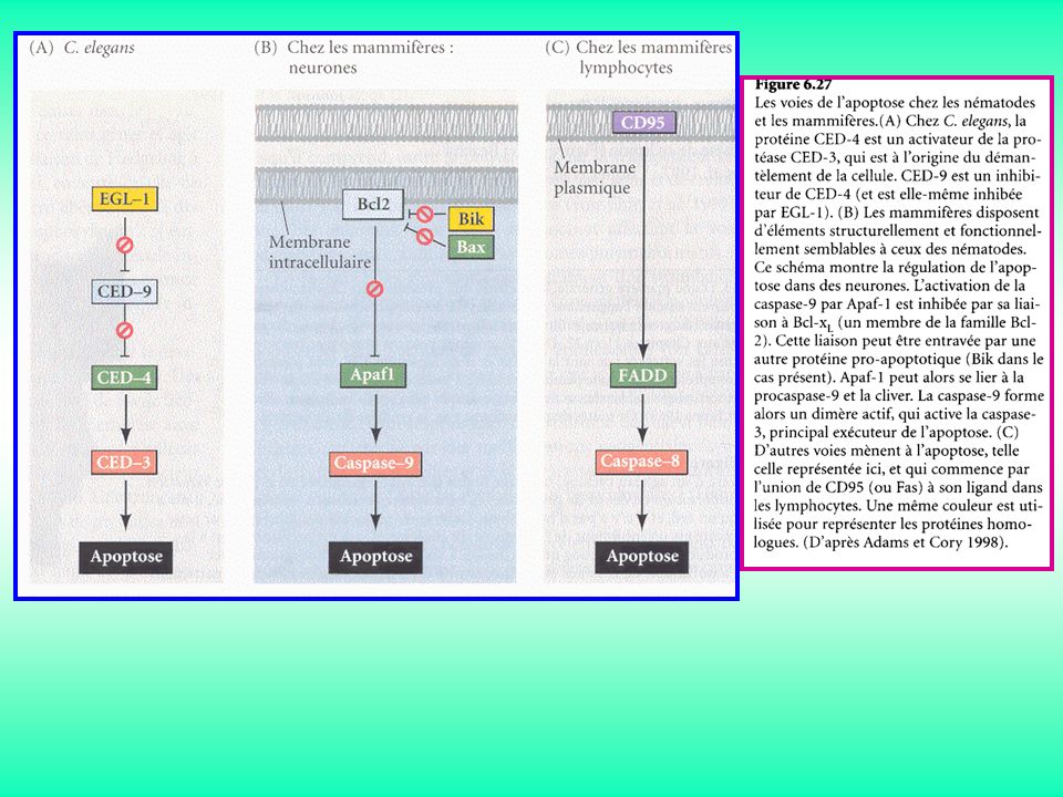
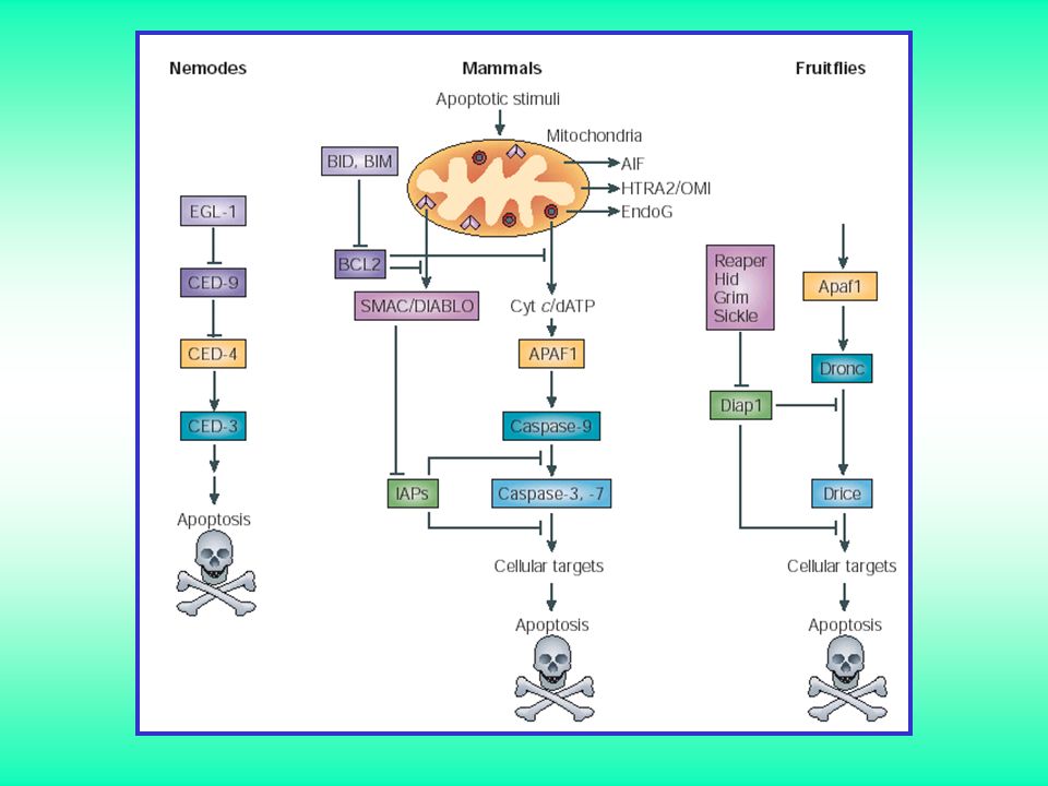
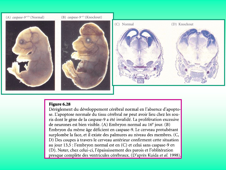
APOPTOSE

60
Figure 3. Résumé schématique des deux principales voies apoptotiques cellulaires. Adapté de Koutouras, J

65
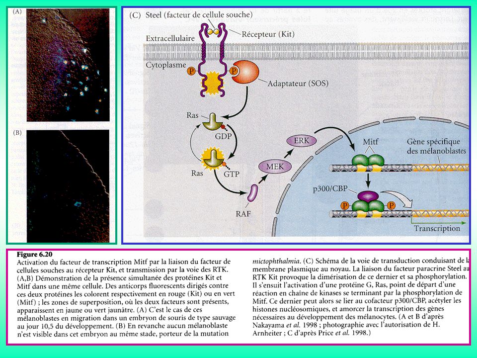
Kit/SCF (Stem Cell Factor)
Bcl-2

66
SIGNALISATION JUXTACRINES

67
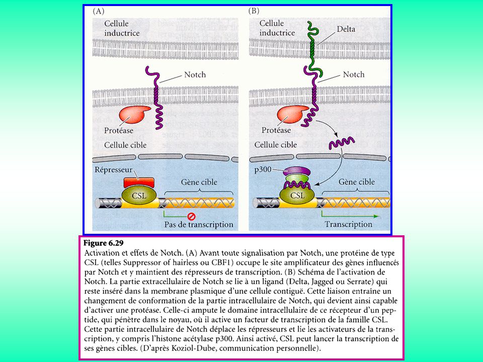
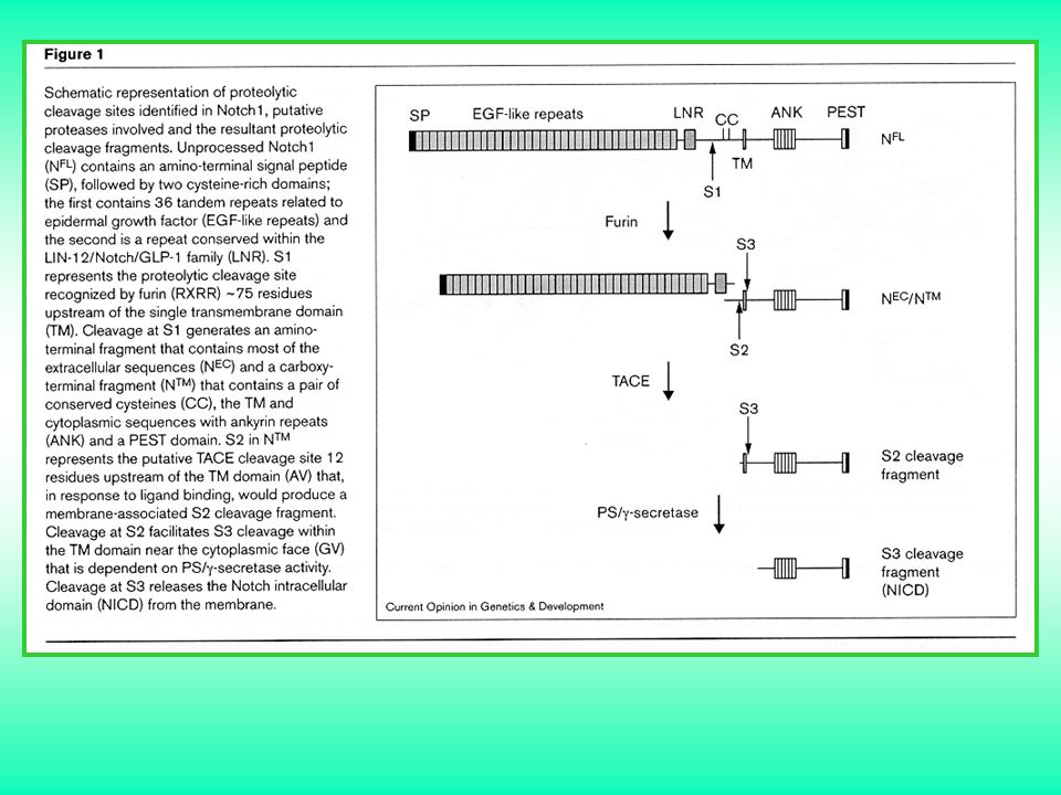
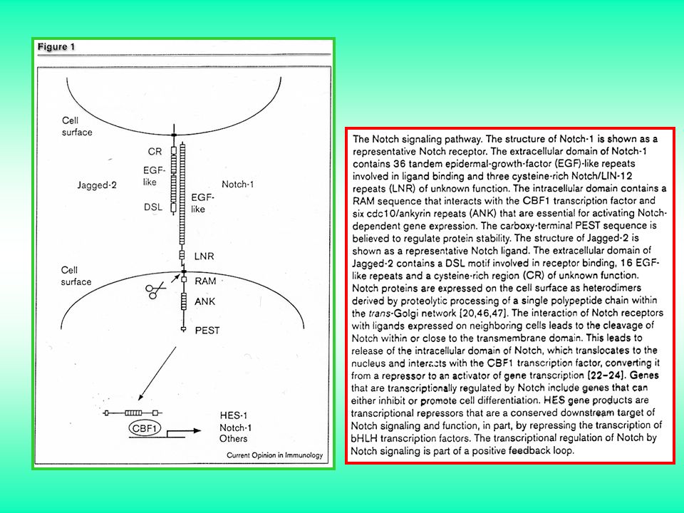
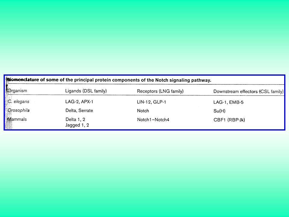
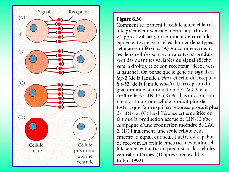
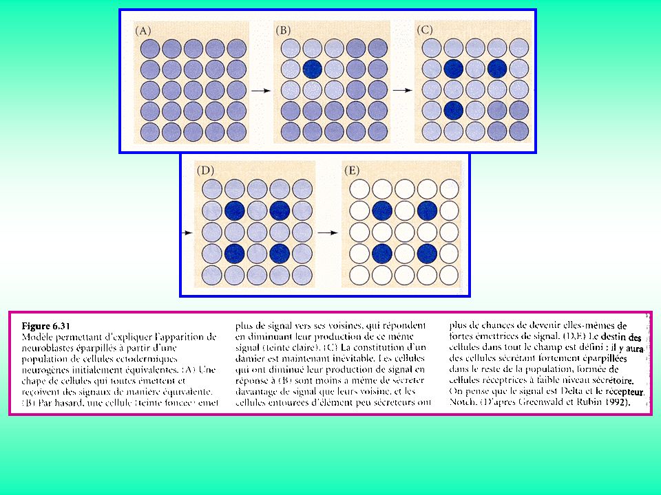
Signalisation Notch

73
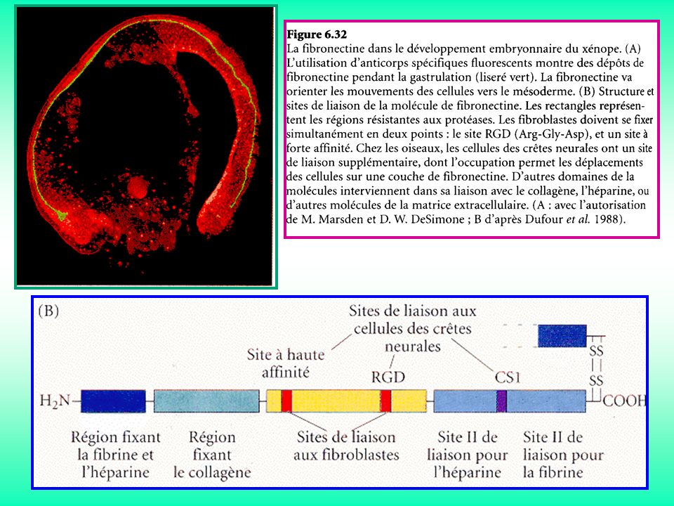
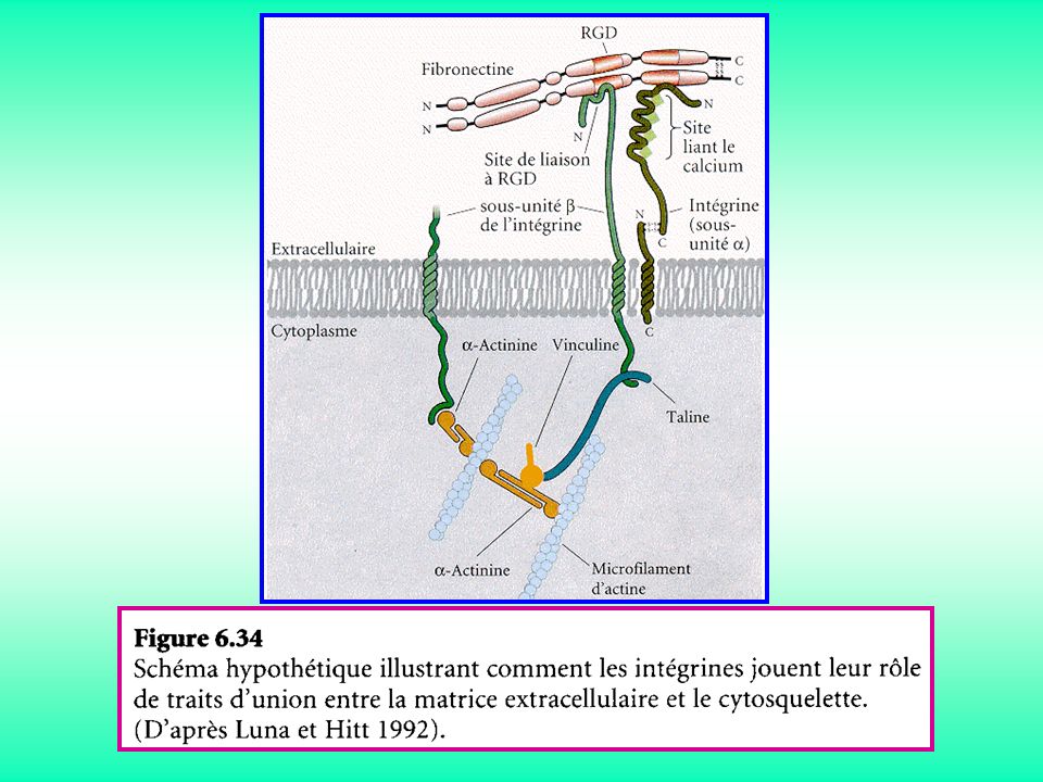
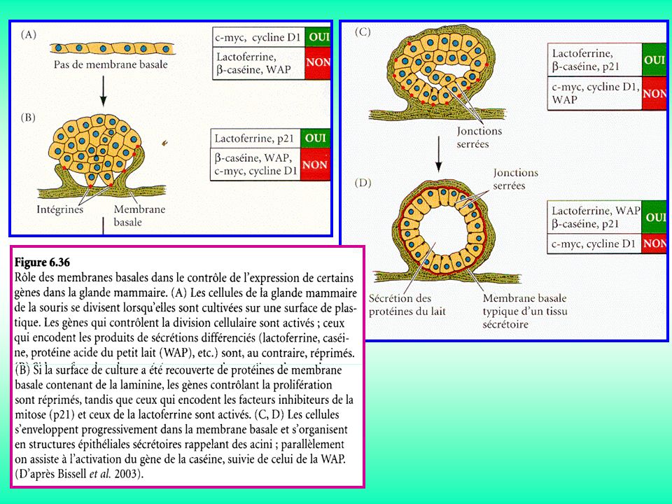
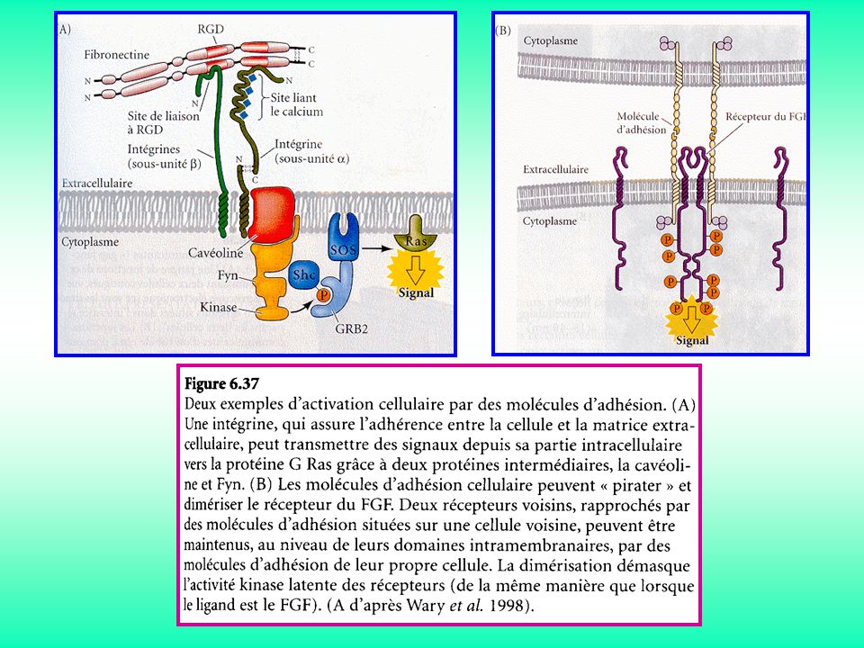
Signalisation ECM

77
(A) (B)
 (B)")
80
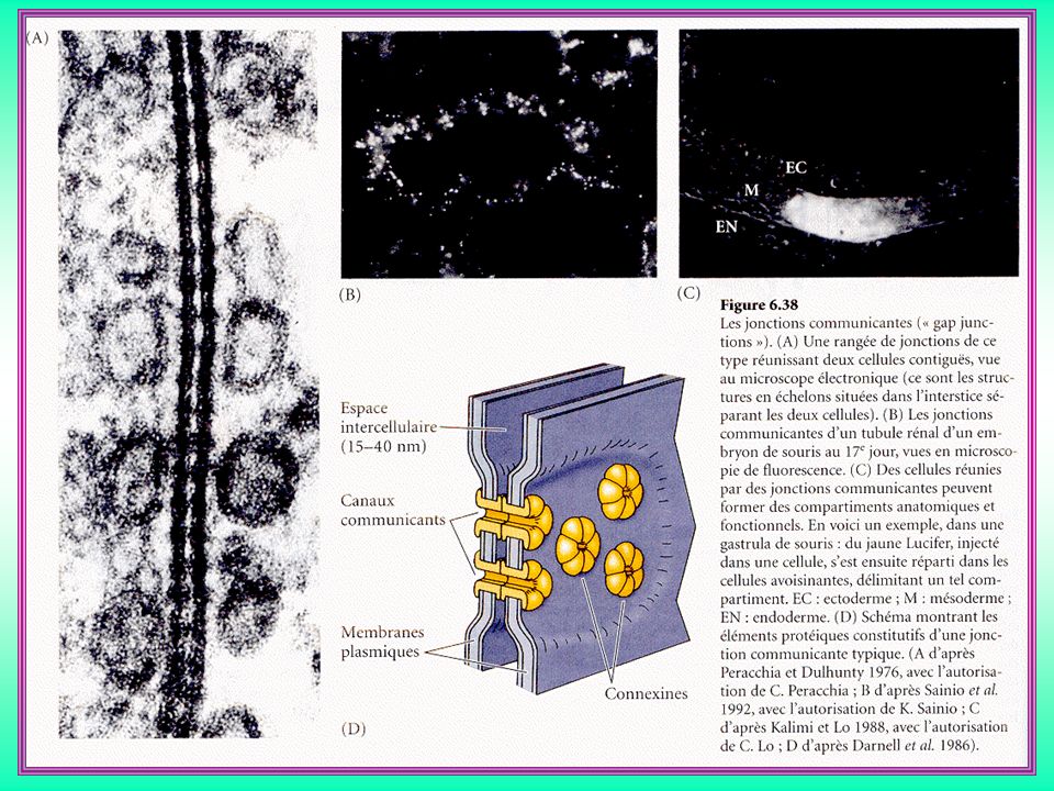
Jonctions gap

82
A B

83
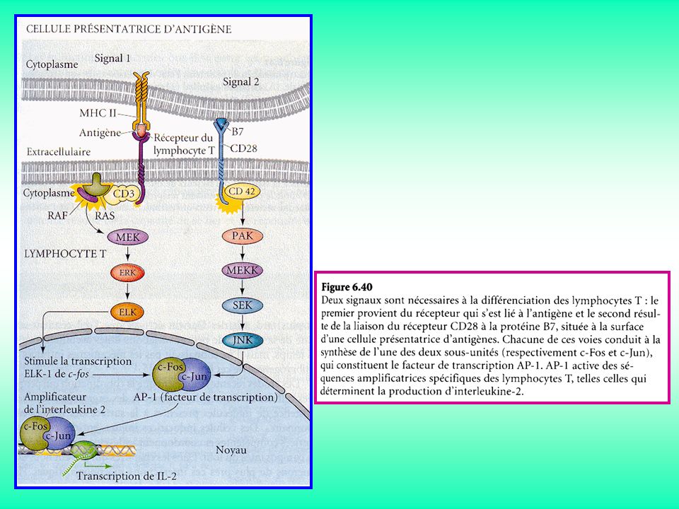
Dialogues entre signalisations

85
Maintient de l’état différencié

87
Détermination cellulaire :
hasard et dialogues intercellulaire

Présentations similaires