Télécharger la présentation
La présentation est en train de télécharger. S'il vous plaît, attendez

1
Saignement Gastro-intestinal
Samah Chiry, R1

2
Objectifs 1. Chez un patient qui a du sang dans ses selles et dont l’état hémodynamique est instable, tenez compte des antécédents pour distinguer entre un saignement gastro-intestinal supérieur et un saignement inférieur, car l’examen sera alors différent. 2. Chez un patient chez qui vous soupçonnez la présence de sang dans les selles, explorez d’autres causes possibles (p. ex. ingestion de betteraves, de fer, de Pepto-Bismol) avant de procéder à un examen général. 3. Recherchez les patients à risque plus élevé de saignement GI (p. ex. saignement GI antérieur, séjour aux soins intensifs, anti-inflammatoires non stéroïdiens, alcool) afin de modifier le traitement pour diminuer le risque de saignement GI (p. ex. cytoprotection) 4. Chez les patients souffrant d’un saignement GI évident, identifiez ceux qui pourraient nécessiter un traitement en temps opportun même s’ils ne sont pas encore en état de choc. 5. Chez un patient stable atteint d’un saignement GI inférieur, recherchez des causes sérieuses (p. ex. tumeur, maladie intestinale inflammatoire, ulcère, varices) même quand le saignement semble avoir une cause évidente (p. ex. n’attribuez pas un saignement rectal aux hémorroïdes ou à un anticoagulant oral). 6. Chez un patient souffrant d’un saignement GI supérieur : a) incluez le saignement de varices dans votre diagnostic différentiel; b) utilisez les antécédents et l’examen physique pour évaluer la probabilité d’un saignement variqueux, car sa prise en charge est différente.
 avant de procéder à un examen général. 3. Recherchez les patients à risque plus élevé de saignement GI (p. ex. saignement GI antérieur, séjour aux soins intensifs, anti-inflammatoires non stéroïdiens, alcool) afin de modifier le traitement pour diminuer le risque de saignement GI (p. ex. cytoprotection) 4. Chez les patients souffrant d’un saignement GI évident, identifiez ceux qui pourraient nécessiter un traitement en temps opportun même s’ils ne sont pas encore en état de choc. 5. Chez un patient stable atteint d’un saignement GI inférieur, recherchez des causes sérieuses (p. ex. tumeur, maladie intestinale inflammatoire, ulcère, varices) même quand le saignement semble avoir une cause évidente (p. ex. n’attribuez pas un saignement rectal aux hémorroïdes ou à un anticoagulant oral). 6. Chez un patient souffrant d’un saignement GI supérieur : a) incluez le saignement de varices dans votre diagnostic différentiel; b) utilisez les antécédents et l’examen physique pour évaluer la probabilité d’un saignement variqueux, car sa prise en charge est différente.")
3
Anatomie saignement GI superieur vs inferieur determine par le ligament de treitz (LT). Superieur: proximal au LT: Oesophage, estomac, 2eme / 3eme portion du duodenum inferieur : distal au LT: petit intestin et colon

4
Saignement GI superieur Saignement GI inferieur
Incidence : cas/ adultes / annee Mortalite : 10 a 14% La moyenne pour le sejour a l’hopital (n’incluant pas les varices) avec ou sans complications est de 2.7 a 4.4 jours. Mortalite a diminue de 23% aux EU (1998 a 2006), de 40% en Bretagne (1993 a 2007), inchange au Canada (1993 a 2003) 20-33% des episodes de saignement GI Mortalite: 10 a 20% surtout avec les patients ages et ceux avec comorbidites Plus frequent chez les personnes agees a cause de la haute incidence de la diverticulose et la maladie vasculaire chez ce groupe. ♂ > ♀ Ceci est relie a l’incidence de l’ulcere peptique de l’estomac qui est relie a l’utilisation des NSAIDS ou ASA
 avec ou sans complications est de 2.7 a 4.4 jours. Mortalite a diminue de 23% aux EU (1998 a 2006), de 40% en Bretagne (1993 a 2007), inchange au Canada (1993 a 2003) 20-33% des episodes de saignement GI. Mortalite: 10 a 20% surtout avec les patients ages et ceux avec comorbidites. Plus frequent chez les personnes agees a cause de la haute incidence de la diverticulose et la maladie vasculaire chez ce groupe. ♂ > ♀ Ceci est relie a l’incidence de l’ulcere peptique de l’estomac qui est relie a l’utilisation des NSAIDS ou ASA.")
5
Saignement aigu: moins de trois jours Instabilite hemodynamique et/ou besoin de transfusion sanguine

6
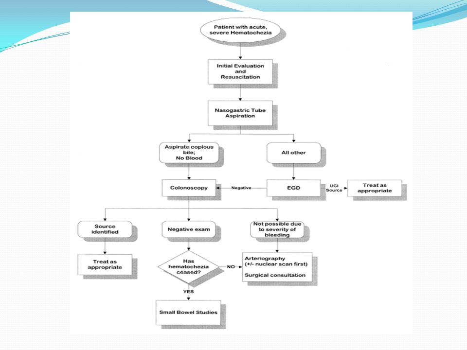
Est ce que le saignement GI est superieur ou inferieur?
Evaluation initiale Le premier point dans la prise en charge d’un saignement digestif est de definir le site et la cause du saignement: Est ce que le saignement GI est superieur ou inferieur?

7
L’histoire La nature du saignement :
Hematemese (vx du sang): origine superieure Coffee ground emesis Rouge frais Caillots de sang Apres des vomissements excessifs Melena Selles noires (verifier avec le TR) A besoin d’au moins 50cc de sang dans le tractus GI Typiquement survient d’une origine superieure, mais 5% peuvent etre d’origine inferieure Occasionnellement la source est le petit intestin ou le colon proximal Hematoschezia (sang rouge per rectum) d’habitude suggere un saignement distal au LT A considerer un saignement superieur si instabilite hemodynamique The color of the vomitus depends on its contact time with the hydrochloric acid of the stomach. If vomiting occurs early after the onset of bleeding, it appears red; with delayed vomiting, it is dark red, brown, or black. a contact time of blood in the gut for 8 hours is required for melena and patients with rapid bleeding from an upper GI source pass bright red blood rectally because of rapid GI transit, About 30% of patients with bleeding ulcers present with hematemesis, 20% with melena, and 50% with both
: origine superieure. Coffee ground emesis. Rouge frais. Caillots de sang. Apres des vomissements excessifs. 2. Melena. Selles noires (verifier avec le TR) A besoin d’au moins 50cc de sang dans le tractus GI. Typiquement survient d’une origine superieure, mais 5% peuvent etre d’origine inferieure. Occasionnellement la source est le petit intestin ou le colon proximal. 3. Hematoschezia (sang rouge per rectum) d’habitude suggere un saignement distal au LT. A considerer un saignement superieur si instabilite hemodynamique. The color of the vomitus depends on its contact time with the hydrochloric acid of the stomach. If vomiting occurs early after the onset of bleeding, it appears red; with delayed vomiting, it is dark red, brown, or black. a contact time of blood in the gut for 8 hours is required for melena and patients with rapid bleeding from an upper GI source pass bright red blood rectally because of rapid GI transit, About 30% of patients with bleeding ulcers present with hematemesis, 20% with melena, and 50% with both.")
8
Les points importants dans l’histoire
La duree du saignement, la frequence Les sx associes: douleur abd, un nouveau chgt dans les habitudes de selles, fievre, urgence/tenesme, perte de poids, anorexie, N/V, satiete precoce, dysphagie/odynophagie, epistaxis/hemoptysie Antcds: Saignement anterieur Trauma Hx de radiation a l’abd et au pelvis Ulcere peptique anterieur Hx de maladie inflammatoire de l’intestin Comorbidites : MCAS, ins cardiaque, maladie hepatique Chx anterieure: AAA Endoscopies anterieures : les rapports Medication: NSAIDs ASA Anticoagulants

9
Laboratoire FSC Base : E, Cr, Uree
(uree peut augmenter sans augmentation de la Cr du a l’absorption des proteines du sang et a la dehydratation) PT/PTT Type et crossmatch ECG chez les patients > 50 ans, ou ayant une hx de MCAS ou arrythmie, ou ayant une DRS/palpitations associees avec le saignement Never rely on initial H/H values to asses amount of blood loss (hemoconcentration), diminue dramatiquement apres l’hydratation However une absence d’augmentation de l’uree ne r/o pas UGIB.
 PT/PTT. Type et crossmatch. ECG chez les patients > 50 ans, ou ayant une hx de MCAS ou arrythmie, ou ayant une DRS/palpitations associees avec le saignement. Never rely on initial H/H values to asses amount of blood loss (hemoconcentration), diminue dramatiquement apres l’hydratation. However une absence d’augmentation de l’uree ne r/o pas UGIB.")
10
Diagnostic differentiel : saignement GI superieur
Variceal bleeding, a severe outcome of portal hypertension secondary to cirrhosis, carries a 6-week mortality rate of 10% to 20%.50 In view of the risk, primary prevention is indicated in patients with high-risk varices A Dieulafoy erosion is an abnormal cirsoid aneurysmal artery that protrudes through a tiny mucosal defect, usually within 6 cm of the gastroesophageal junction on the lesser curve of the stomach. Les causes rares: ulcere de stress,ulcere de Dieulafoy, malformation AV, Fistule aorto-enterique, anastomose chirurgicale..

11
Sx de la maladie hepatique
Saignement variqueux Sx de la maladie hepatique Antecedants Faiblesse, anorexie et malaise Nausee et vomissement Perte de poids Discomfort ou douleur abdominal (QSD, epigastre et QSG) Jaunisse et urine foncee Oedeme et ascite Prurit Ecchymoses (easy bruising) Impotence et dysfonction sexuel Crampes musculaires Hx de jaunisse suggerant une hepatite anterieure infectieuse ou medicamenteuse Hx de transfusion Hx de drogues IV Prise d’alcool ou AINS Hx de cirrhose All variceal bleeding should be considered severe, since the 1-year death rate is so high (up to 70%). Clues pointing to variceal bleeding include previous variceal bleeding, thrombocytopenia, history of liver disease, and signs of liver disease on clinical examination. All patients suspected of having bleeding varices should be admitted to the intensive care unit for close monitoring and should be given the highest priority, even if they are hemodynamically stable
 Jaunisse et urine foncee. Oedeme et ascite. Prurit. Ecchymoses (easy bruising) Impotence et dysfonction sexuel. Crampes musculaires. Hx de jaunisse suggerant une hepatite anterieure infectieuse ou medicamenteuse. Hx de transfusion. Hx de drogues IV. Prise d’alcool ou AINS. Hx de cirrhose. All variceal bleeding should be considered severe, since the 1-year death rate is so high (up to 70%). Clues pointing to variceal bleeding include previous variceal bleeding, thrombocytopenia, history of liver disease, and signs of liver disease on clinical examination. All patients suspected of having bleeding varices should be admitted to the intensive care unit for close monitoring and should be given the highest priority, even if they are hemodynamically stable.")
12
Saignement variqueux Paleur
Hypertrophie des parotides (reliee a l’alcolisme et/ou malnutrition) Jaunisse Telangiectasis ( peau, levres, doigts). Gynecomastie chez les hommes Fetor hepaticus erytheme palmaire Ascites Circulation collaterale autour de l’ombilic, venous hums (bruit audible du flot turbulent dans les veines collaterales) Slenomegalie Atrophie testiculaire Angiome stellare Caput medusa hemorrhoides
 Jaunisse. Telangiectasis ( peau, levres, doigts). Gynecomastie chez les hommes. Fetor hepaticus. erytheme palmaire. Ascites. Circulation collaterale autour de l’ombilic, venous hums (bruit audible du flot turbulent dans les veines collaterales) Slenomegalie. Atrophie testiculaire. Angiome stellare. Caput medusa. hemorrhoides.")
13
Diagnostic differentiel : saignement GI inferieur
Sites du petit intestin : ileite de crohn, meckel’s diverticulum, ectasie vasculaire

14
Recommendations de la prise en charge des patients avec saignement GI superieur non variqueux
A. Resuscitation, risk assessment, and preendoscopy management A1. Immediately evaluate and initiate appropriate resuscitation. A2. Prognostic scales are recommended for early stratification of patients into low- and high-risk categories for rebleeding and mortality.† A3. Consider placement of a nasogastric tube in selected patients because the findings may have prognostic value.* A4. Blood transfusions should be administered to a patient with a hemoglobin level 70 g/L. A5. In patients receiving anticoagulants, correction of coagulopathy is recommended but should not delay endoscopy. A6. Promotility agents should not be used routinely before endoscopy to increase the diagnostic yield. A7. Selected patients with acute ulcer bleeding who are at low risk for rebleeding on the basis of clinical and endoscopic criteria may be discharged promptly after endoscopy.† A8. Preendoscopic PPI therapy may be considered to downstage the endoscopic lesion and decrease the need for endoscopic intervention but should not delay

15
Determiner l’urgence de la situation clinique:
Est ce que le patient est en choc? Tachypnee, tachycardie, agitation, diminution de l’etat de conscience, palleur indiquent une perte de sang > 1500 mL ( 30% du volume circulatoire) Att aux beta-bloqueurs 2. Est ce que le patient est orthostatique? SV avec TA couche et debout Une ↓ de > 10 mmhg ou une ↑ de > 10 batt/min indiquent une perte de sang > 800 mL (15% du volume circulatoire)
 Att aux beta-bloqueurs. 2. Est ce que le patient est orthostatique SV avec TA couche et debout. Une ↓ de > 10 mmhg ou une ↑ de > 10 batt/min indiquent une perte de sang > 800 mL (15% du volume circulatoire)")
16
Recommendations de la prise en charge des patients avec saignement GI superieur non variqueux
A. Resuscitation, risk assessment, and preendoscopy management A1. Immediately evaluate and initiate appropriate resuscitation. A2. Prognostic scales are recommended for early stratification of patients into low- and high-risk categories for rebleeding and mortality. A3. Consider placement of a nasogastric tube in selected patients because the findings may have prognostic value.* A4. Blood transfusions should be administered to a patient with a hemoglobin level 70 g/L. A5. In patients receiving anticoagulants, correction of coagulopathy is recommended but should not delay endoscopy. A6. Promotility agents should not be used routinely before endoscopy to increase the diagnostic yield. A7. Selected patients with acute ulcer bleeding who are at low risk for rebleeding on the basis of clinical and endoscopic criteria may be discharged promptly after endoscopy.† A8. Preendoscopic PPI therapy may be considered to downstage the endoscopic lesion and decrease the need for endoscopic intervention but should not delay

17
Les predicteurs cliniques pour resaignement ou mortalite
Age > 65 ans Choc Comorbidites Hb initialement bas Melena Besoin de transfusion Sang rouge frais au TR, dans le Vx, ou le TNG Sepsis ↑ uree, creat, AST, ALT

18
Glasgow- Blatchford Bleeding score
Uree (mmol/L) 6·5-7·9 = 2 points 8·0-9·9 = 3 points 10·0-25·0 = 4 points >25·0 = 6 points Haemoglobin pour les hommes (g/L) =1 points = 3 points <100 = 6 points Haemoglobin pour les femmes (g/L) =1 point <100 = 6 point Pression systolique(mm Hg) =1point 90-99 =2 point <90 =3 point D’autres marqueurs: Pouls ≥ 100/min =1 point presentation avec melena = 1 point presentation avec syncope = 2 point Maladie hepatique= 2 point Insuffisance cardique=2 point Ceux qui ont un score de 0 peuvent avoir leur conge sans endoscopie
 6·5-7·9 = 2 points. 8·0-9·9 = 3 points. 10·0-25·0 = 4 points. >25·0 = 6 points. Haemoglobin pour les hommes (g/L) =1 points = 3 points. <100 = 6 points. Haemoglobin pour les femmes (g/L) =1 point. <100 = 6 point. Pression systolique(mm Hg) =1point =2 point. <90 =3 point. D’autres marqueurs: Pouls ≥ 100/min =1 point. presentation avec melena = 1 point. presentation avec syncope = 2 point. Maladie hepatique= 2 point. Insuffisance cardique=2 point. Ceux qui ont un score de 0 peuvent avoir leur conge sans endoscopie.")
19
Rockall scoring system
the most widely used, gives estimates of the risks of recurrent bleeding and death. It is based on the three clinical factors mentioned above and on two endoscopic ones, awarding points for: Age—0 points if less than 60; 1 point if 60 to 79; or 2 points if 80 years or older Shock—1 point if the pulse is more than 100; 2 points if the systolic blood pressure is less than 100 mm Hg Comorbid illness—2 points for ischemic heart disease, congestive heart failure, or other major comorbidity; 3 points for renal failure, hepatic failure, or metastatic disease Endoscopic diagnosis—0 points if no lesion found or a Mallory-Weiss tear; 1 point for peptic ulcer, esophagitis, or erosive disease; 2 points for GI malignancy Endoscopic stigmata or recent hemorrhage—0 points for a clean-based ulcer or flat pigmented spot; 2 points for blood in the upper GI tract, active bleeding, a nonbleeding visible vessel, or adherent clot. The Rockall score can thus range from 0 to 11 points, with an overall score of 0, 1, or 2 associated with an excellent prognosis.

20
Recommendations de la prise en charge des patients avec saignement GI superieur non variqueux
A. Resuscitation, risk assessment, and preendoscopy management A1. Immediately evaluate and initiate appropriate resuscitation. A2. Prognostic scales are recommended for early stratification of patients into low- and high-risk categories for rebleeding and mortality. A3. Consider placement of a nasogastric tube in selected patients because the findings may have prognostic value. A4. Blood transfusions should be administered to a patient with a hemoglobin level 70 g/L. A5. In patients receiving anticoagulants, correction of coagulopathy is recommended but should not delay endoscopy. A6. Promotility agents should not be used routinely before endoscopy to increase the diagnostic yield. A7. Selected patients with acute ulcer bleeding who are at low risk for rebleeding on the basis of clinical and endoscopic criteria may be discharged promptly after endoscopy.† A8. Preendoscopic PPI therapy may be considered to downstage the endoscopic lesion and decrease the need for endoscopic intervention but should not delay

21
Tube NasoGastrique Indication :
Devant tout saignement superieur ou saignement indetermine Benefices therapeutiques et diagnostiques Positive evidence que le saignement est superieur sang rouge frais endoscopie en urgence coffee ground endoscopie ds prochains 24h Negative n’exclut pas un saignement superieur (saignement intermittent, spasme pylorique, oedeme empechant le reflux du sang duodenal) Contraindications: Varices oesophagiennes ne sont pas une contraindication Trauma facial/ fracture de l’os nasal
 Contraindications: Varices oesophagiennes ne sont pas une contraindication. Trauma facial/ fracture de l’os nasal.")
22
Recommendations de la prise en charge des patients avec saignement GI superieur non variqueux
A. Resuscitation, risk assessment, and preendoscopy management A1. Immediately evaluate and initiate appropriate resuscitation. A2. Prognostic scales are recommended for early stratification of patients into low- and high-risk categories for rebleeding and mortality. A3. Consider placement of a nasogastric tube in selected patients because the findings may have prognostic value. A4. Blood transfusions should be administered to a patient with a hemoglobin level 70 g/L. A5. In patients receiving anticoagulants, correction of coagulopathy is recommended but should not delay endoscopy. A6. Promotility agents should not be used routinely before endoscopy to increase the diagnostic yield. A7. Selected patients with acute ulcer bleeding who are at low risk for rebleeding on the basis of clinical and endoscopic criteria may be discharged promptly after endoscopy.† A8. Preendoscopic PPI therapy may be considered to downstage the endoscopic lesion and decrease the need for endoscopic intervention but should not delay

23
Recommendations de la prise en charge des patients avec saignement GI superieur non variqueux
A. Resuscitation, risk assessment, and preendoscopy management A1. Immediately evaluate and initiate appropriate resuscitation. A2. Prognostic scales are recommended for early stratification of patients into low- and high-risk categories for rebleeding and mortality. A3. Consider placement of a nasogastric tube in selected patients because the findings may have prognostic value. A4. Blood transfusions should be administered to a patient with a hemoglobin level 70 g/L. A5. In patients receiving anticoagulants, correction of coagulopathy is recommended but should not delay endoscopy. A6. Promotility agents should not be used routinely before endoscopy to increase the diagnostic yield. A7. Selected patients with acute ulcer bleeding who are at low risk for rebleeding on the basis of clinical and endoscopic criteria may be discharged promptly after endoscopy.† A8. Preendoscopic PPI therapy may be considered to downstage the endoscopic lesion and decrease the need for endoscopic intervention but should not delay The number of people presenting with upper GI bleeding while on warfarin therapy is increasing because of the expanding indications for long-term anticoagulation therapy, such as atrial fibrillation and deep venous thrombosis. The risk of GI bleeding in patients who use oral anticoagulants is estimated to be 2.3 to 4.9 times higher than in nonusers.61 The goal international normalized ratio (INR) for patients on warfarin therapy is usually 2.0 to 3.0. Recent studies found that endoscopy can be safely performed in patients with acute GI bleeding whose INR is between 2.0 and ,63 Some suggest that both the length of warfarin therapy and the INR affect the risk of bleeding.64,65 Managing patients with an INR higher than 3.0 who have an episode of GI bleeding is always a challenge. It is not uncommon to find pathologic lesions causing GI bleeding in patients who are on warfarin with a supratherapeutic INR, and thus, endoscopy is indicated. However, before endoscopy, reversal of anticoagulation should be considered.
 for patients on warfarin therapy is usually 2.0 to 3.0. Recent studies found that endoscopy can be safely performed in patients with acute GI bleeding whose INR is between 2.0 and ,63 Some suggest that both the length of warfarin therapy and the INR affect the risk of bleeding.64,65. Managing patients with an INR higher than 3.0 who have an episode of GI bleeding is always a challenge. It is not uncommon to find pathologic lesions causing GI bleeding in patients who are on warfarin with a supratherapeutic INR, and thus, endoscopy is indicated. However, before endoscopy, reversal of anticoagulation should be considered.")
24
Warfarin peut precipite un saignement
Le risque de saignement GI chez les patients ayant du coumadin est de 2.3 a 4.9 fois plus grand que ceux qui n’ont pas. Malgre que le nbre de plaquettes n’est pas un predicteur de la mortalite, un INR > 1.5 a la presentation est un predicteur significatif de la mortalite chez les patients avec saignement superieur. Endoscopie peut se faire avec un INR entre 2-3 Si INR supratherapeutique, il faut le corriger avant.

25
Recommendations de la prise en charge des patients avec saignement GI superieur non variqueux
A. Resuscitation, risk assessment, and preendoscopy management A1. Immediately evaluate and initiate appropriate resuscitation. A2. Prognostic scales are recommended for early stratification of patients into low- and high-risk categories for rebleeding and mortality. A3. Consider placement of a nasogastric tube in selected patients because the findings may have prognostic value. A4. Blood transfusions should be administered to a patient with a hemoglobin level 70 g/L. A5. In patients receiving anticoagulants, correction of coagulopathy is recommended but should not delay endoscopy. A6. Promotility agents should not be used routinely before endoscopy to increase the diagnostic yield. A7. Selected patients with acute ulcer bleeding who are at low risk for rebleeding on the basis of clinical and endoscopic criteria may be discharged promptly after endoscopy.† A8. Preendoscopic PPI therapy may be considered to downstage the endoscopic lesion and decrease the need for endoscopic intervention but should not delay endoscopy.

26
Recommendations de la prise en charge des patients avec saignement GI superieur non variqueux
B. Endoscopic management B1. Develop institution-specific protocols for multidisciplinary management. Include access to an endoscopist trained in endoscopic hemostasis. B2. Have available on an urgent basis support staff trained to assist in endoscopy. B3. Early endoscopy (within 24 hours of presentation) is recommended for most patients with acute upper gastrointestinal bleeding. B4. Endoscopic hemostatic therapy is not indicated for patients with low-risk stigmata (a clean-based ulcer or a nonprotuberant pigmented dot in an ulcer bed). B5. A finding of a clot in an ulcer bed warrants targeted irrigation in an attempt at dislodgement, with appropriate treatment of the underlying lesion. B6. The role of endoscopic therapy for ulcers with adherent clots is controversial. Endoscopic therapy may be considered, although intensive PPI therapy alone may be sufficient. B7. Endoscopic hemostatic therapy is indicated for patients with high-risk stigmata (active bleeding or a visible vessel in an ulcer bed). B8. Epinephrine injection alone provides suboptimal efficacy and should be used in combination with another method. B9. No single method of endoscopic thermal coaptive therapy is superior to another. B10. Clips, thermocoagulation, or sclerosant injection should be used in patients with high-risk lesions, alone or in combination with epinephrine injection. B11. Routine second-look endoscopy is not recommended. B12. A second attempt at endoscopic therapy is generally recommended in cases of rebleeding.
 is recommended for most patients with acute upper gastrointestinal bleeding. B4. Endoscopic hemostatic therapy is not indicated for patients with low-risk stigmata (a clean-based ulcer or a nonprotuberant pigmented dot in an ulcer bed). B5. A finding of a clot in an ulcer bed warrants targeted irrigation in an attempt at dislodgement, with appropriate treatment of the underlying lesion. B6. The role of endoscopic therapy for ulcers with adherent clots is controversial. Endoscopic therapy may be considered, although intensive PPI therapy alone may be sufficient. B7. Endoscopic hemostatic therapy is indicated for patients with high-risk stigmata (active bleeding or a visible vessel in an ulcer bed). B8. Epinephrine injection alone provides suboptimal efficacy and should be used in combination with another method. B9. No single method of endoscopic thermal coaptive therapy is superior to another. B10. Clips, thermocoagulation, or sclerosant injection should be used in patients with high-risk lesions, alone or in combination with epinephrine injection. B11. Routine second-look endoscopy is not recommended. B12. A second attempt at endoscopic therapy is generally recommended in cases of rebleeding.")
27
Recommendations de la prise en charge des patients avec saignement GI superieur non variqueux
Pharmacologic management C1. Histamine-2 receptor antagonists are not recommended for patients with acute ulcer bleeding. C2. Somatostatin and octreotide are not routinely recommended for patients with acute ulcer bleeding. C3. An intravenous bolus followed by continuous-infusion PPI therapy should be used to decrease rebleeding and mortality in patients with high-risk stigmata who have undergone successful endoscopic therapy. C4. Patients should be discharged with a prescription for a single daily-dose oral PPI for a duration as dictated by the underlying etiology. these drugs did not prove very useful in managing upper GI bleeding in clinical trials.17,18 In their intravenous form, they often fail to keep the gastric pH at 6 or higher, due to tachyphylaxis

28
IPP La dose recommendee d’omeprazole: 80 mg bolus suivi d’une perfusion de 8 mg/h pour 72 h diminue le resaignement , l’indication pour la chirurgie et la mortalite. Apres le traitement de l’evenement aigu, un IPP est recommende pour 6 a 8 semaines, plus long si test positif pour H.pylori ou s’il prend l’ASA ou un AINS.

29
Algorithm for patients with acute upper gastrointestinal bleeding.
ALBELDAWI M et al. Cleveland Clinic Journal of Medicine 2010;77: ©2010 by Cleveland Clinic

30
Endoscopic stigmata of bleeding peptic ulcer (arrows) and risk of recurrent bleeding and death. Upper endoscopy is 90% to 95% diagnostic for acute upper GI bleeding. Endoscopic stigmata of bleeding peptic ulcer (arrows) and risk of recurrent bleeding and death. ALBELDAWI M et al. Cleveland Clinic Journal of Medicine 2010;77: ©2010 by Cleveland Clinic
 and risk of recurrent bleeding and death. ALBELDAWI M et al. Cleveland Clinic Journal of Medicine 2010;77: ©2010 by Cleveland Clinic.")
31
Recommendations de la prise en charge des patients avec saignement GI superieur non variqueux
D. Nonendoscopic and nonpharmacologic in-hospital management D1. Patients at low risk after endoscopy can be fed within 24 hours. D2. Most patients who have undergone endoscopic hemostasis for high-risk stigmata should be hospitalized for at least 72 hours thereafter. D3. Seek surgical consultation for patients for whom endoscopic therapy has failed. D4. Where available, percutaneous embolization can be considered as an alternative to surgery for patients for whom endoscopic therapy has failed. D5. Patients with bleeding peptic ulcers should be tested for H. pylori and receive eradication therapy if it is present, with confirmation of eradication. D6. Negative H. pylori diagnostic tests obtained in the acute setting should be repeated. Les etudes ont montre que les lesions a haut risque ont besoin de 72h apres trt endoscopique pour devenir des lesions a bas risque

32
Preferred therapies for Helicobacter pylori infection
Triple therapy : Proton pump inhibitor twice a day + Clarithromycin (Biaxin) 500 mg twice a day + Amoxicillin 1,000 mg twice a day –14 days 70%–85% Proton pump inhibitor twice a day + Clarithromycin 500 mg twice a day + Metronidazole (Flagyl) 500 mg twice a day 10–14 days 75%–85% Consider in penicillin-allergic patients who have not previously received a macrolide or who cannot tolerate bismuth quadruple therapy Quadruple therapy Proton pump inhibitor twice a day + Bismuth subsalicylate 525 mg twice a day + Metronidazole 250 mg four times a day + Tetracycline 500 mg four times a day 75%–90% Consider in penicillin-allergic patients
 500 mg twice a day + Amoxicillin 1,000 mg twice a day. - 10–14 days. 70%–85% Proton pump inhibitor twice a day + Clarithromycin 500 mg twice a day + Metronidazole (Flagyl) 500 mg twice a day. 10–14 days. 75%–85% Consider in penicillin-allergic patients who have not previously received a macrolide or who cannot tolerate bismuth quadruple therapy. Quadruple therapy. Proton pump inhibitor twice a day + Bismuth subsalicylate 525 mg twice a day + Metronidazole 250 mg four times a day + Tetracycline 500 mg four times a day. 75%–90% Consider in penicillin-allergic patients.")
33
Recommendations de la prise en charge des patients avec saignement GI superieur non variqueux
E. Postdischarge, ASA, and NSAIDs E1. In patients with previous ulcer bleeding who require an NSAID, it should be recognized that treatment with a traditional NSAID plus PPI or a COX-2 inhibitor alone is still associated with a clinically important risk for recurrent ulcer bleeding. E2. In patients with previous ulcer bleeding who require an NSAID, the combination of a PPI and a COX-2 inhibitor is recommended to reduce the risk for recurrent bleeding from that of COX-2 inhibitors alone. E3. In patients who receive low-dose ASA and develop acute ulcer bleeding, ASA therapy should be restarted as soon as the risk for cardiovascular complication is thought to outweigh the risk for bleeding. E4. In patients with previous ulcer bleeding who require cardiovascular prophylaxis, it should be recognized that clopidogrel alone has a higher risk for rebleeding than ASA combined with a PPI.

34
ASA The higher the aspirin dose, the higher the risk. Aspirin doubles the risk of upper GI bleeding at daily doses of 75 mg , and quadruples it at doses of 300 mg. The most important risk factor for upper GI bleeding in patients taking aspirin is a history of peptic ulcer bleeding. Approximately 15% of aspirin users who have bleeding from ulcers have recurrent bleeding within 1 year. When considering whether to discontinue antiplatelet therapy, a cardiologist should be consulted along with a gastroenterologist to weigh the risks of GI bleeding vs thrombosis.

35
Clopidogrel (Plavix) Clopidogrel (Plavix) is recommended for hospitalized patients with acute coronary syndrome who cannot tolerate the GI side effects of aspirin. This recommendation was largely based on the safety data from the CAPRIE (Clopidogrel Versus Aspirin in Patients at Risk of Ischemic Events) trial, in which the incidence of major GI bleeding was lower in the clopidogrel group (0.52%) than in the aspirin group (0.72%; P < .05).
 is recommended for hospitalized patients with acute coronary syndrome who cannot tolerate the GI side effects of aspirin. This recommendation was largely based on the safety data from the CAPRIE (Clopidogrel Versus Aspirin in Patients at Risk of Ischemic Events) trial, in which the incidence of major GI bleeding was lower in the clopidogrel group (0.52%) than in the aspirin group (0.72%; P < .05).")
36
In a recent double-blind randomized controlled trial in patients with a history of aspirin-induced bleeding, the combination of low-dose aspirin plus esomeprazole (Nexium) twice a day was superior to clopidogrel by itself in terms of the rate of recurrent bleeding (0.7% vs 8.6%; P < .05). A similar trial showed nearly identical results: 0% upper GI bleeding in the group receiving aspirin plus esomeprazole 20 mg daily, vs 13.6% in the clopidogrel group (P = .0019). These studies suggest that a once-daily proton pump inhibitor combined with aspirin is a safer alternative than clopidogrel alone
. These studies suggest that a once-daily proton pump inhibitor combined with aspirin is a safer alternative than clopidogrel alone.")
37
Clopidogrel plus a proton pump inhibitor
Interestingly, recent studies have shown that omeprazole (Losec) decreases the antiplatelet effect of clopidogrel, possibly by inhibiting the CYP2C19 enzyme. However, concomitant use of , lansoprazole (Prevacid), and esomeprazole (Nexium) did not have this effect, suggesting that although all proton pump inhibitors are metabolized to a varying degree by CYP2C19, the interaction between proton pump inhibitors and clopidogrel is not a class effect. Therefore, pantoprazole, lansoprazole, and esomeprazole may be the appropriate proton pump inhibitors to use with clopidogrel in patients who have a clear indication for the medication, consistent with current guideline recommendations.
 decreases the antiplatelet effect of clopidogrel, possibly by inhibiting the CYP2C19 enzyme. However, concomitant use of , lansoprazole (Prevacid), and esomeprazole (Nexium) did not have this effect, suggesting that although all proton pump inhibitors are metabolized to a varying degree by CYP2C19, the interaction between proton pump inhibitors and clopidogrel is not a class effect. Therefore, pantoprazole, lansoprazole, and esomeprazole may be the appropriate proton pump inhibitors to use with clopidogrel in patients who have a clear indication for the medication, consistent with current guideline recommendations.")
39
Indications pour la chirurgie
Emergency surgery is required in about 10-25% of patients with lower gastrointestinal bleeding (LGIB). The indications for surgery : 1. Persistent hemodynamic instability with active bleeding 2. Persistent, recurrent bleeding 3. Transfusion of more than 4 units packed red bloods cells in a 24-hour period, with active or recurrent bleeding
. The indications for surgery : 1. Persistent hemodynamic instability with active bleeding. 2. Persistent, recurrent bleeding. 3. Transfusion of more than 4 units packed red bloods cells in a 24-hour period, with active or recurrent bleeding.")
41
Take Home message The first priority is to ensure that the patient is hemodynamically stable, which often requires admission to the intensive care unit for monitoring and fluid resuscitation. Peptic ulcers account for most cases of upper GI bleeding, but bleeding from varices has a much higher case-fatality rate and always demands aggressive treatment. Patients with ulcer disease should be tested and treated for Helicobacter pylori infection. Patients with a history of bleeding ulcers who need long-term treatment with aspirin or a nonsteroidal anti-inflammatory drug should also be prescribed a proton pump inhibitor.

42
Bibliographie International Consensus Recommendations on the Management of Patients With Nonvariceal Upper Gastrointestinal Bleeding, Annals of Internal Medicine, 2010; 152: Management of the Adult Patient With Acute Lower Gastrointestinal Bleeding, American Journal of Gastroenterology (1998) 93, 1202–1208
 93, 1202–1208.")
Présentations similaires


>")


