Télécharger la présentation
La présentation est en train de télécharger. S'il vous plaît, attendez

1
FRACTURES DU CALCANEUM
1 2 A.MENADI UNIVERSITE-BADJI MOKHTAR-ANNABA FACULTE DE MEDECINE DEPARTEMENT DE MEDECINE

2
INTRODUCTION Les fractures du calcanéum sont définies comme une solution de continuité de l’os calcanéen. Ce sont des fractures graves, car la plupart d’entre elles touchent la région articulaire(thalamique) Le traitement de ces fractures *orthopédiquement, *chirurgicalement * fonctionnel reste le traitement de base. Il est souvent utilisé seul ; sinon il doit être associé à d’autres moyens thérapeutiques.
 Le traitement de ces fractures. *orthopédiquement, *chirurgicalement * fonctionnel reste le traitement de base. Il est souvent utilisé seul ; sinon il doit être associé à d’autres moyens thérapeutiques.")
3
RAPPEL ANATOMIQUE ET PHYSIOLOGIQUE
1 1-Calcanéum –astragale:arrière pied 2- 6 faces: -supérieure:post,ant thalamus+++ -inf:peau épaisse -Ant:articulaire cuboide -post:insertion tendon achille -Int:sustentaculum tali -ext:chirugicale 3-APPUI MONO PODAL il supporte 2/3 poids corps 4-mvts: inversion-éversion

4
ETIOLOGIES-MECANISMES
1-chute lieu élevé+++ (acc trav-suicide-acc domestique…. 2-MECANISMES:INDIRECT *CISSAILLEMENT: 2 forces :sol -poids corps>>>calcan coupé en 2 *COMPRESSION: THALAMUS VERTICALE -HORIZONTALE 1 2 1 2

5
ANATOMIE PATHOLOGIQUE
*LESIONS ELEMENTAIRES: -SEPARATION:fr séparation,trait fr oblique,calcanéum séparée 2 parties post ext-antero interne. -ENFONCEMENT:fr COMPRESSION, fr enfoncement vertical, mécanisme flexion dorsale fr enfoncement horizontal, mécanisme flexion plantaire 1 2 1 2

6
ANATOMIE PATHOLOGIQUE classification DUPARC
1-FR EXTRA THALAMIQUES: A-fr tubérosité post:- fr A° POST SUP(bec canard),Achille++ -fr tub post inf,court fp,add 1 -fr deux tubér B-fr grande apophyse:art calc cuboid C-fr sustentaculum tali 1 2 1 2
,Achille++ -fr tub post inf,court fp,add 1. -fr deux tubér. B-fr grande apophyse:art calc cuboid. C-fr sustentaculum tali")
7
ANATOMIE PATHOLOGIQUE classification DUPARC
2-FR THALAMIQUES:5 types *type 1:fr séparation 2 fragment,trait oblique *type 2:fr luxation,partie post int luxe dehors *type 3:fr séparation-compression a 3 fragments -partie antéro interne -partie post externe,(fr tubéros+fr thalamique) 1 2 1 2
")
8
ANATOMIE PATHOLOGIQUE classification DUPARC
*fr type 3 degré 1:verticalisation modéré,angle BOEHLER + *fr type 3 degré 2:verticalisation GRANDE,angle BOEHLER nul *fr type 3 degré 3:verticalisation très importante,angle BOEHLER --- fr type 3 degré 1:enfoncement modéré,angle BOEHLER + *fr type 3 degré 2:enfocement GRAND,angle BOEHLER nul *fr type 3 degré 3:enfoncement très important,angle BOEHLER --- 1 2

9
ANATOMIE PATHOLOGIQUE classification DUPARC
*type 4:fr séparation compression a 4 fragments,fr corticale plantaire,4° FRAGMENT tuberos post ext *Type 5: comminutive 1 2 1 2

10
ANATOMIE PATHOLOGIQUE LESIONS ASSOCIES
1-lesions cutanées: fr type 5,CDB-GUST 2-lesions VASCULO NERVEUSES: rare 3-lesions OSSEUSES:fractures rachis dorso lombaire chute lieu élevée,RX systématique

11
ETUDE CLINIQUE 1-INTERROGATOIRE:heure accident-mecanismes-age-antcd-tares 2-EXAMEN PHYSIQUE: *oedéme arriére pied, disparition sillons, *EXAMEN CUTANEE: classification CDB-GUST *EXAMEN VASCULAIRE: artère tibial post 4P GRIFFITH *EXAMEN OSTEO ARTICULAIRE:PILON TIBIAL-CALCANEUM- MALLEO et surtout rachis lombaire+++ 1 2 3

12
EXAMEN RADIOGRAPHIQUE
1-radios(05)- face –profil-rot int-rot ext-rétro tibiale 2-angle BOEHLER:
- face –profil-rot int-rot ext-rétro tibiale. 2-angle BOEHLER:")
13
EXAMEN RADIOGRAPHIQUE
1-radios(05)- face –profil-rot int-rot ext-rétro tibiale 2-angle BOEHLER:25-40° -ligne sommet grande tubérosité -ligne sommet thalamus 3-incidence rétro tibiale: -visualiser grande tubérosité, -surface thalamique -différencier type 3 ou 4 1 2
- face –profil-rot int-rot ext-rétro tibiale. 2-angle BOEHLER:25-40° -ligne sommet grande tubérosité. -ligne sommet thalamus. 3-incidence rétro tibiale: -visualiser grande tubérosité, -surface thalamique. -différencier type 3 ou")
14
EXAMEN RADIOGRAPHIQUE
4- tomodensitométrie( scanner) -étudie déplacement,enfoncement
 -étudie déplacement,enfoncement.")
15
EVOLUTION-COMPLICATIONS
1-DELAI DE CSLD: SEMAINES 2-complications immédiates: *cutanées:ecchymose ,plaie cutanée *nerveuses -vasculaires:rare *osseuses:fr rachis lombaires+++++ 3-complicatrions TARDIVES *cal vicieux thalamique ou extra thalamique *ARTHROSE arriére pied *osteite chronique: secondaire fr ouverte

16
TRAITEMENT 1-BUT:obtenir arrière pied fonctionnel 2-MOYENS:
*fonctionnelles :(fr type 5) -3 phases:1repos-réeducation-2marche sans appui-3appui *orthopédiques:(fr parcellaire,fr type 1 ) -botte platrée GRAFFIN( appui plantaire) *chirurgicales:(type ) Matériels:vissage-embrochage,plaque -ostéosynthése foyer fermée - -ostéosynthése foyer ouvert
 -3 phases:1repos-réeducation-2marche sans appui-3appui. *orthopédiques:(fr parcellaire,fr type 1 ) -botte platrée GRAFFIN( appui plantaire) *chirurgicales:(type ) Matériels:vissage-embrochage,plaque. -ostéosynthése foyer fermée. - -ostéosynthése foyer ouvert.")
17
TRAITEMENT ostéosynthése foyer fermée
1 2

18
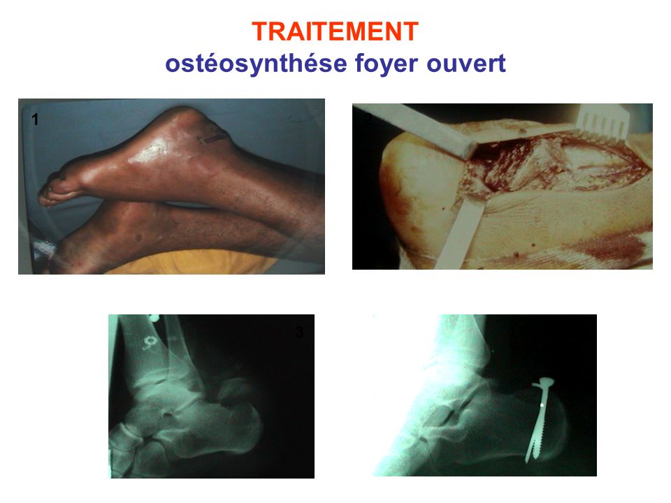
TRAITEMENT ostéosynthése foyer ouvert
1 2 3 4
Présentations similaires


