Télécharger la présentation
La présentation est en train de télécharger. S'il vous plaît, attendez

1
Le monde Microbien Bactéries (taille 1-5 µm) monocellulaire
Procaryotes (noyau simplifié, paroi) Multiplication rapide sur milieux (109 <24h) Virus (taille 10 nm) Un seul acide nucleïque (ADN ou ARN) Multiplication dans cellules obligatoire Prions ou Agent Non Conventionnel Proteine, accumulation intracellulaire Parasites-Champignons eucaryotes monocellulaire Protozoaires (Paludisme..) ou pluricellulaires: helminthes - Arthropodes
 Multiplication rapide sur milieux (109 <24h) Virus (taille 10 nm) Un seul acide nucleïque (ADN ou ARN) Multiplication dans cellules obligatoire. Prions ou Agent Non Conventionnel. Proteine, accumulation intracellulaire. Parasites-Champignons eucaryotes. monocellulaire Protozoaires (Paludisme..) ou pluricellulaires: helminthes - Arthropodes.")
2
Existence de micro-organismes responsables d'infection
Epidémies et moyens de lutte Tableaux cliniques identiques Transmission entre individus (Peste 25 millions de morts 1347 en Europe) Infection expérimentale Agent infectieux responsable Reproductibilité (Postulats de Koch 1884) Microscope optique microscope 17ème s, coloration de Gram fin 19 ème s Microscopie électronique Mise en évidence Virus (agents ultrafiltrables à travers filtres bactériens 0,25 μm)
 Infection expérimentale. Agent infectieux responsable. Reproductibilité (Postulats de Koch 1884) Microscope optique. microscope 17ème s, coloration de Gram fin 19 ème s. Microscopie électronique. Mise en évidence Virus (agents ultrafiltrables à travers filtres bactériens 0,25 μm)")
3
Prions ou Agents Transmissibles Non Conventionnels
Accumulation dans le système nerveux – encéphalopathie spongiforme Grande résistance aux protocoles de base de désinfection

4
Virus à symétrie hélicoïdale et cubique

5
Microscopie électronique
Bactéries Microscopie électronique Microscopie optique Coloration de Gram

6
Champignons Forme levure Forme filamenteuse

7
Protozoaires

8
Ectoparasites

9
Taille des agents infectieux

10
Epidémies 20ème siècle Tuberculose Paludisme Amibiase Schistosomiase
Filariose Rougeole HIV SIDA Poliomyélite 19ème siècle Variole Choléra Diphtérie Lèpre Tuberculose Typhoide Amérique du sud, Afrique et Asie Europe

11
Culture des Bactéries “Génération spontanée?” (Pasteur 1861)
Chirurgie antiseptique (Lister 1867) Lavage mains et réduction fièvre puerpérale (Ignace Semmelweis 1847) Agents ultra-filtrables-Microscopie électronique
 Lavage mains et réduction fièvre puerpérale (Ignace Semmelweis 1847) Agents ultra-filtrables-Microscopie électronique.")
12
La majorité des micro-organismes ne sont pas pathogènes: notion de niche écologique
Homme: les flores Cutanée tube digestif autres muqueuses Eaux douces, mer, résiduaires sols propres et contaminés Végétaux Animaux

13
Notion de Pathogénicité
germes saprophytes Danger 0 Bénéfice Hôte (symbiose) germes commensaux Danger +/- Bénéfice Hôte +/- (mutualisme) germes pathogènes Danger + Bénéfice parasite (parasitisme) Flore « normale » ou flore de barrière à préserver Limiter au maximum le danger de la flore commensale Prévenir le contact ou la susceptibilité aux germes pathogènes
 germes commensaux. Danger +/- Bénéfice Hôte +/- (mutualisme) germes pathogènes. Danger + Bénéfice parasite (parasitisme) Flore « normale » ou flore de barrière à préserver. Limiter au maximum le danger de la flore commensale. Prévenir le contact ou la susceptibilité aux germes pathogènes.")
14
Comment les micro-organismes peuvent ils être responsable d'infection?
Facteurs de virulence Adhérence (infection urinaire) Toxines (Choléra) autres facteurs limitant les défenses de l’organisme ou favorisant la multiplication des germes.
 Toxines (Choléra) autres facteurs limitant les défenses de l’organisme ou favorisant la multiplication des germes.")
15
Bactéries Flagelles Membrane externe Peptidoglycan (endotoxine)
Plasmide Chromosome Adhésines

16
La microscopie électronique montre la structure type prokaryote
The higher resolving power of the electron microscope not only magnifies the typical shape of a bacterial cell but also clearly resolves its prokaryotic organization FIGURE 2-2 Electron micrograph of a thin section of Neisseria gonorrhoeae showing the organizational features of prokaryotic cells. Note the electron-transparent nuclear region (n) packed with DNA fibrils, the dense distribution of ribosomal particles in the cytoplasm, and the absence of intracellular membranous organelles. The Nucleoid Prokaryotic and eukaryotic cells were initially distinguished on the basis of structure: the prokaryotic nucleoidthe equivalent of the eukaryotic nucleusis structurally simpler than the true eukaryotic nucleus, which has a complex mitotic apparatus and surrounding nuclear membrane. As the electron micrograph in Fig. 2-2 shows, the bacterial nucleoid, which contains the DNA fibrils, lacks a limiting membrane. Under the light microscope, the nucleoid of the bacterial cell can be visualized with the aid of Feulgen staining, which stains DNA. Gentle lysis can be used to isolate the nucleoid of most bacterial cells.
 packed with DNA fibrils, the dense distribution of ribosomal particles in the cytoplasm, and the absence of intracellular membranous organelles. The Nucleoid. Prokaryotic and eukaryotic cells were initially distinguished on the basis of structure: the prokaryotic nucleoidthe equivalent of the eukaryotic nucleusis structurally simpler than the true eukaryotic nucleus, which has a complex mitotic apparatus and surrounding nuclear membrane. As the electron micrograph in Fig. 2-2 shows, the bacterial nucleoid, which contains the DNA fibrils, lacks a limiting membrane. Under the light microscope, the nucleoid of the bacterial cell can be visualized with the aid of Feulgen staining, which stains DNA. Gentle lysis can be used to isolate the nucleoid of most bacterial cells.")
17
Prokaryotes et Eucaryotes ont une organisation cellulaire différentes
Prokaryotes have a nucleoid (nuclear body) rather than an enveloped nucleus and lack membrane-bound cytoplasmic organelles. The plasma membrane in prokaryotes performs many of the functions carried out by membranous organelles in eukaryotes. Multiplication is by binary fission.
 rather than an enveloped nucleus and lack membrane-bound cytoplasmic organelles. The plasma membrane in prokaryotes performs many of the functions carried out by membranous organelles in eukaryotes. Multiplication is by binary fission.")
18
Présence d ’appendices externes caractéristiques: pili (fimbriae) et flagelles
Surface Structures FIGURE 2-3 (A) Electron micrograph of negatively stained E coli showing wavy flagella and numerous short, thinner, and more rigid hairlike structures, the pili. (B) The long sex pilus can be distinguished from the shorter common pili by mixing E coli cells with a male bacteriophage that binds specifically to sex pili. Flagella: The flagella of motile bacteria differ in structure from eukaryotic flagella. A basal body anchored in the plasma membrane and cell wall gives rise to a cylindrical protein filament. The flagellum moves by whirling about its long axis. The number and arrangement of flagella on the cell are diagnostically useful. Pili (Fimbriae): Pili are slender, hairlike, proteinaceous appendages on the surface of many (particularly Gram-negative) bacteria. They are important in adhesion to host surfaces. Capsules: Some bacteria form a thick outer capsule of high-molecular-weight, viscous polysaccharide gel; others have more amorphous slime layers. Capsules confer resistance to phagocytosis.
 Electron micrograph of negatively stained E coli showing wavy flagella and numerous short, thinner, and more rigid hairlike structures, the pili. (B) The long sex pilus can be distinguished from the shorter common pili by mixing E coli cells with a male bacteriophage that binds specifically to sex pili. Flagella: The flagella of motile bacteria differ in structure from eukaryotic flagella. A basal body anchored in the plasma membrane and cell wall gives rise to a cylindrical protein filament. The flagellum moves by whirling about its long axis. The number and arrangement of flagella on the cell are diagnostically useful. Pili (Fimbriae): Pili are slender, hairlike, proteinaceous appendages on the surface of many (particularly Gram-negative) bacteria. They are important in adhesion to host surfaces. Capsules: Some bacteria form a thick outer capsule of high-molecular-weight, viscous polysaccharide gel; others have more amorphous slime layers. Capsules confer resistance to phagocytosis.")
19
La microscopie électronique révèle des différences anatomiques importantes entre Bactéries Gram + et Gram - FIGURE 2-5 (A) Electron micrograph of a thin section of the Gram-positive M lysodeikticus showing the thick peptidoglycan cell wall (cw), underlying cytoplasmic (plasma) membrane (cm), mesome (m), and nucleus (n). (B) Freeze-fractured Bacteriodes cell showing typical major convex fracture faces through the inner (im) and outer (om) membranes. Bars = 1 µm; circled arrow in Fig. B indicates direction of shadowing. The topographic relationships of the cell wall and envelope layers to the plasma membrane are indicated in the thin section of a Gram-positive organism (Micrococcus lysodeikticus) in Figure 2-5A and in the freeze-fractured cell of a Gram-negative organism (Bacteroides melaninogenicus) in Figure 2-5B. The latter shows the typical fracture planes seen in most Gram-negative bacteria, which are weak cleavage planes through the outer membrane of the envelope and extensive fracture planes through the bilayer region of the underlying plasma membrane.
 Electron micrograph of a thin section of the Gram-positive M lysodeikticus showing the thick peptidoglycan cell wall (cw), underlying cytoplasmic (plasma) membrane (cm), mesome (m), and nucleus (n). (B) Freeze-fractured Bacteriodes cell showing typical major convex fracture faces through the inner (im) and outer (om) membranes. Bars = 1 µm; circled arrow in Fig. B indicates direction of shadowing. The topographic relationships of the cell wall and envelope layers to the plasma membrane are indicated in the thin section of a Gram-positive organism (Micrococcus lysodeikticus) in Figure 2-5A and in the freeze-fractured cell of a Gram-negative organism (Bacteroides melaninogenicus) in Figure 2-5B. The latter shows the typical fracture planes seen in most Gram-negative bacteria, which are weak cleavage planes through the outer membrane of the envelope and extensive fracture planes through the bilayer region of the underlying plasma membrane.")
20
Classification des bactéries
Morphologie et coloration de Gram Coques, bacilles, spirilles… Gram+, Gram-, autres…. Caractéres culturaux Aérobie, anaérobie, microaérophiles Exigences nutritionnelles Profil biochimique Génétique Pathogénicité

21
Classification

22
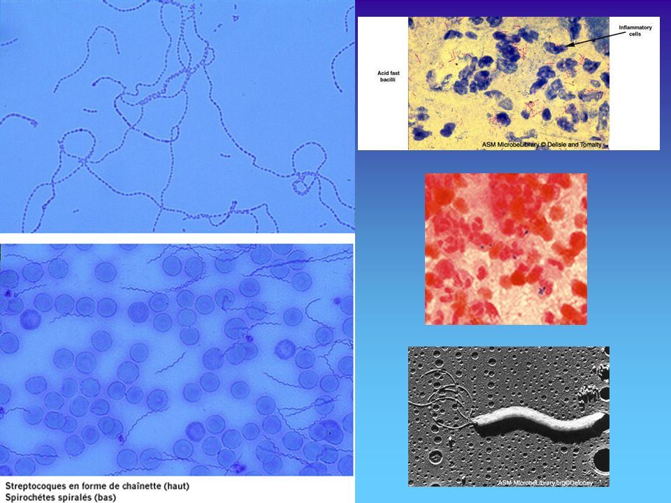
Morphologie et colonisation

24
Différentiation des colonies

25
Pourquoi tous les individus ne font pas une infection?
Immunité naturelle Immunité acquise les moyens de défense de l'organisme Mécaniques humoraux immunité cellulaire non spécifique immunité cellulaire spécifique Autres facteurs: fièvre-cytokines Nutrition

26
Immunité cellulaire non spécifique

27
Bactéries intra-cellulaires

28
Adhérence

29
Capsule et phagocytose: Streptococcus pneumoniae

30
Epidémiologie des infections
Prévention Hygiène-vaccination-Prévention transmission Traitement Nouveaux agents pathogènes Adaptation- résistance aux antibiotiques = anciens agents nouveaux agents

Présentations similaires