Télécharger la présentation
La présentation est en train de télécharger. S'il vous plaît, attendez

1
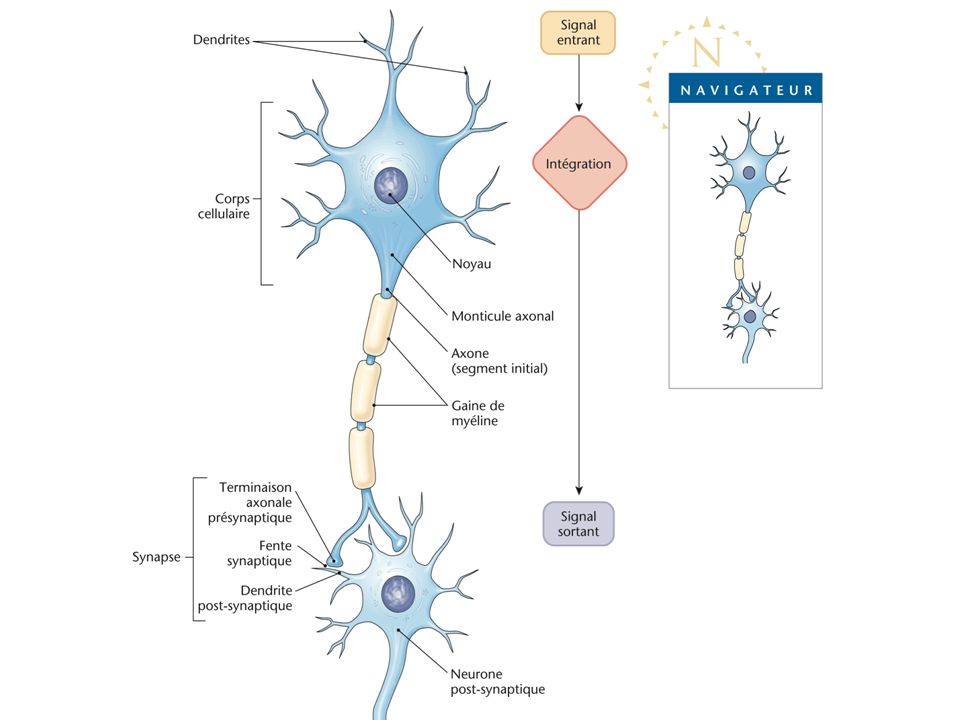
Synapse & message nerveux
Pr Anh Tuan DINH-XUAN Service de Physiologie-Explorations Fonctionnelles, Hôpital Cochin, Faculté de Médecine, Université Paris Descartes

3
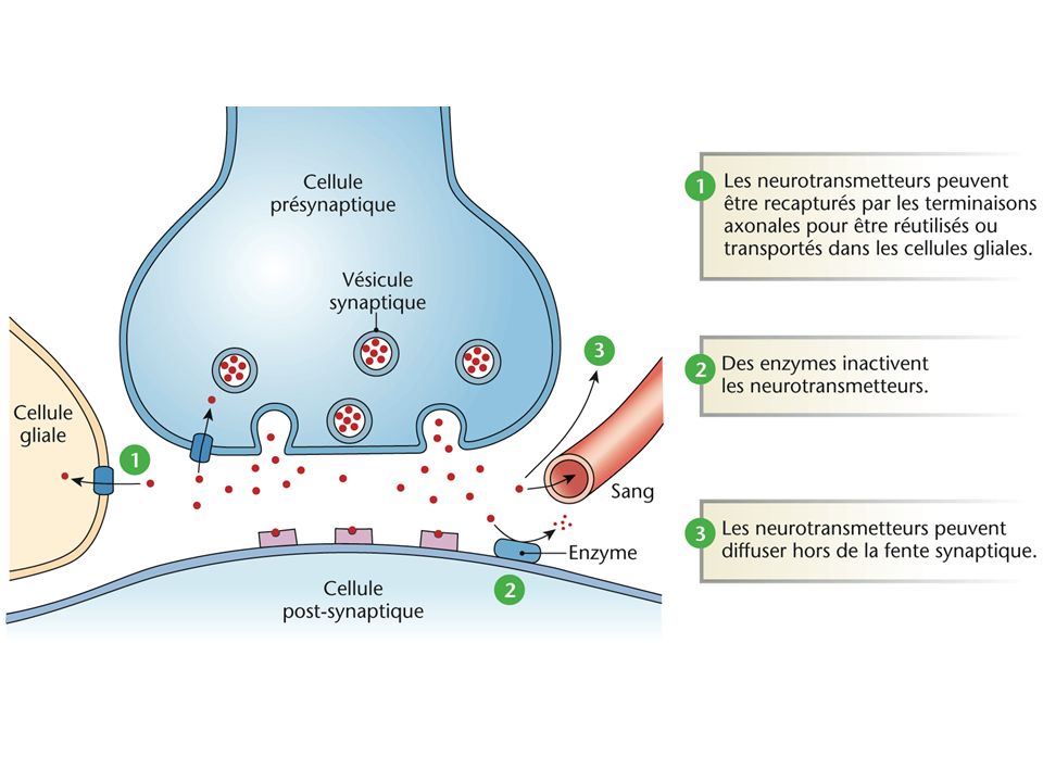
Transmission synaptique

4
Transmission synaptique
Pr AT DINH-XUAN UE8-TC6

5
Transmission synaptique
PPSI PPSE La liaison du neurotransmetteur au récepteur membranaire de la cellule post-synaptique va créer un potentiel post-synaptique (PPS) excitateur (PPSE) ou inhibiteur (PPSI). Transmission synaptique Pr AT DINH-XUAN Figure An EPSP and IPSP (as recorded on an oscilloscope). Each can be generated separately, at the axon hillock, by selective stimulation of an excitatory (E) or inhibitory (I) input to a neurone. If the two occur so that they overlap then they add at the axon hillock. K+ Cl- Na+ Ca2+ [-90 à –65 mV] UE8-TC6
 excitateur (PPSE) ou inhibiteur (PPSI). Transmission synaptique. Pr AT DINH-XUAN. Figure An EPSP and IPSP (as recorded on an oscilloscope). Each can be generated separately, at the axon hillock, by selective stimulation of an excitatory (E) or inhibitory (I) input to a neurone. If the two occur so that they overlap then they add at the axon hillock. K+ Cl- Na+ Ca2+ [-90 à –65 mV] UE8-TC6.")
6
Neurotransmetteurs : classification
Les neurotransmetteurs sont divisés en deux principaux groupes : les petites molécules les peptides

7
Neurotransmetteurs (petites molécules)
Neurotransmetteurs dérivés des acides aminés : Monoamines Catécholamines Autres Purines (ATP) Gaz (NO, CO) PETITES MOLECULES Acétylcholine Monoxyde d’azote Monoxyde de carbonne
 Gaz (NO, CO) PETITES MOLECULES. Acétylcholine. Monoxyde d’azote. Monoxyde de carbonne.")
8
Monoamines Figure 12-9 Biosynthesis of some common small transmitter molecules.

9
Catécholamines Noradrénaline Adrénaline
Figure 12-9 Biosynthesis of some common small transmitter molecules. Adrénaline

10
SMALL-MOLECULE NEUROTRANSMITTERS
Amino Acids Glutamate Gamma aminobutyric acid (GABA) Glycine Aspartate Neuro-inhibiteur Neuro-excitateur
 Glycine. Aspartate. Neuro-inhibiteur. Neuro-excitateur.")
11
GABA receptors contain a chloride channel
Chloride ion (Cl–) GABA Plasma membrane I 1. Channel closed until receptor binds to GABA GABA receptors Channel closed 2. GABA receptor binds to GABA, Cl– channel opens 3. Diffusion of Cl– into cell causes hyperpolarization (IPSP)
 GABA. Plasma. membrane. I. 1. Channel closed. until receptor binds. to GABA. GABA. receptors. Channel. closed. 2. GABA receptor. binds to GABA, Cl– channel opens. 3. Diffusion of Cl– into cell causes. hyperpolarization (IPSP)")
12
Transmission synaptique
Pr AT DINH-XUAN Transmission glutamatergique UE8-TC6

13
Neurotransmetteurs (NO)
Figure Nitric oxide (NO) synthesis in a central neuron. Pre-synaptic glutamate release triggers the entry of Ca2+ through NMDA glutamate receptor channels or voltage-gated Ca2+ channels. Via calmodulin (CaM), Ca2+ stimulates nitric oxide synthase (NOS; p. 110). NO diffuses out and through cells to affect presynaptic and postsynaptic elements of the same synapse or of nearby synapses. NADPH, nicotin-amide adenine dinucleotide phosphate; NMDA, N-methyl-d-aspartate.
 synthesis in a central neuron. Pre-synaptic glutamate release triggers the entry of Ca2+ through NMDA glutamate receptor channels or voltage-gated Ca2+ channels. Via calmodulin (CaM), Ca2+ stimulates nitric oxide synthase (NOS; p. 110). NO diffuses out and through cells to affect presynaptic and postsynaptic elements of the same synapse or of nearby synapses. NADPH, nicotin-amide adenine dinucleotide phosphate; NMDA, N-methyl-d-aspartate.")
14
Neuropeptides (neurotransmetteurs & neurohormones)
Figure Structure of some neuroactive peptides. All peptides are presented with their NH2 termini (i.e., the first to be synthesized) to the left, as is now customary for proteins in general. However, note that for many of the peptide hormones, the amino-acid residues were numbered before this convention was established. The p on the amino-terminal glutamate on some of these peptides stands for "pyroglutamate."
 to the left, as is now customary for proteins in general. However, note that for many of the peptide hormones, the amino-acid residues were numbered before this convention was established. The p on the amino-terminal glutamate on some of these peptides stands for pyroglutamate.")
16
Hydrolyse enzymatique
Transporteur membranaire Recapture Elimination Figure The inactivation of acetylcholine. Acetylcholinesterase splits the transmitter, releasing choline which is then recaptured by the nerve and reused in the vesicle to form more acetylcholine. Hydrolyse enzymatique

17
Récepteur pré-synaptique
Figure The release of acetylcholine. Triggered by the action potential, Ca2+ diffuses into the cell, causing a conformational change in the link between the vesicle and the varicosity membranes. Acetylcholine is released and acts on pre- and postjunctional receptors. Récepteur post-synaptique

18
Myasthénie Ptosis Immunobiology Part V. The Immune System in Health and Disease Autoimmunity and Transplantation Autoimmune responses are directed against self antigens. Figure Autoantibodies inhibit receptor function in myasthenia gravis. In normal circumstances, acetylcholine released from stimulated motor neurons at the neuromuscular junction binds to acetylcholine receptors on skeletal muscle cells, triggering muscle contraction (left panel). Myasthenia gravis is caused by autoantibodies against the a subunit of the receptor for acetylcholine. These autoantibodies bind to the receptor without activating it and also cause receptor internalization and degradation (right panel). As the number of receptors on the muscle is decreased, the muscle becomes less responsive to acetylcholine.
. Myasthenia gravis is caused by autoantibodies against the a subunit of the receptor for acetylcholine. These autoantibodies bind to the receptor without activating it and also cause receptor internalization and degradation (right panel). As the number of receptors on the muscle is decreased, the muscle becomes less responsive to acetylcholine.")
19
Alzheimer Disease Associated with loss of cholinergic neurons that synapse on the areas of the brain responsible for memory

20
APP: Amyloid Precursor Protein
sAPP: Soluble extracellular domain of APP Aβ: Amyloid β peptide AICD : Aβ peptide and intracellular C-terminal domain of APP

21
Figure3.Synaptic Dysfunction in Alzheimer’s Disease.
Synaptic loss correlates best with cognitive decline in Alzheimer’s disease. A control synapse is shown at the top of the figure. At the bottom of the figure, an “Alzheimer’s disease synapse” depicting the pleiotropic effects of the .-amyloid peptide (A.) is shown. Rings represent synaptic vesicles. Experimental application and expression of A., especially oligomers, impair synaptic plasticity by altering the balance between long-term potentiation (LTP) and long-term depression (LTD) and reducing the numbers of dendritic spines. At high concentrations, oligomers may suppress basal synaptic transmission. A. facilitates endocytosis of receptors of N-methyl-d-aspartate (NMDAr) and .-amino-3- hydroxy-5-methyl-4-isoxazole propionic acid (AMPAr). A. also binds to the receptors of p75 neurotrophin (p75NTr) and brain-derived neurotrophic factor (the BDNF receptor, also known as the tyrosine kinase B receptor [trkBr]), exacerbating a situation in which levels of BDNF and nerve growth factor (NGF) are already suppressed. A. impairs nicotinic acetylcholine (ACh) receptor (nAChr) signaling and ACh release from the presynaptic terminal. Numbers of hippocampal synapses decrease in mild cognitive impairment in which remaining synaptic profiles show compensatory increases in size. APP denotes amyloid precursor protein, pCaMKII phosphorylated calcium–calmodulin–dependent protein kinase 2, pCREB phosphorylated cyclic AMP response-element-binding protein, trkAr tyrosine kinase A receptor, and VGCC voltage-gated calcium channel.
 is shown. Rings represent synaptic vesicles. Experimental application and expression of A., especially. oligomers, impair synaptic plasticity by altering the balance between long-term potentiation (LTP) and long-term depression (LTD) and reducing the numbers of dendritic spines. At high concentrations, oligomers may suppress basal. synaptic transmission. A. facilitates endocytosis of receptors of N-methyl-d-aspartate (NMDAr) and .-amino-3- hydroxy-5-methyl-4-isoxazole propionic acid (AMPAr). A. also binds to the receptors of p75 neurotrophin (p75NTr) and brain-derived neurotrophic factor (the BDNF receptor, also known as the tyrosine kinase B receptor [trkBr]), exacerbating a situation in which levels of BDNF and nerve growth factor (NGF) are already suppressed. A. impairs. nicotinic acetylcholine (ACh) receptor (nAChr) signaling and ACh release from the presynaptic terminal. Numbers of. hippocampal synapses decrease in mild cognitive impairment in which remaining synaptic profiles show compensatory. increases in size. APP denotes amyloid precursor protein, pCaMKII phosphorylated calcium–calmodulin–dependent protein kinase 2, pCREB phosphorylated cyclic AMP response-element-binding protein, trkAr tyrosine kinase A receptor, and VGCC voltage-gated calcium channel.")
22
Synaptic Plasticity Repeated use of a neuronal pathway may strengthen or reduce synaptic transmission in that pathway. When repeated stimulation enhances excitability, it is called long-term potentiation (LTP). When repeated stimulation reduces excitability, it is called long-term depression (LTD).
. When repeated stimulation reduces excitability, it is called long-term depression (LTD).")
23
N-methyl-d-aspartate receptor (NMDAR)-dependent long-term potentiation (LTP) has been observed in many different brain regions and is dependent on postsynaptic NMDAR activation and calcium/calmodulin-dependent protein-kinase II (CaMKII) for its initiation3. The voltage-dependent relief of the magnesium block of the NMDAR channel allows the synapse to detect coincident presynaptic release of glutamate (Glu) and postsynaptic depolarization. α-amino-3-hydroxy-5-methyl-4-isoxazole propionic acid receptor (AMPAR) insertion into the postsynaptic membrane is a major mechanism underlying LTP expression.
 and postsynaptic depolarization. α-amino-3-hydroxy-5-methyl-4-isoxazole propionic acid receptor (AMPAR) insertion into the postsynaptic membrane is a major mechanism underlying LTP expression..")
24
Presynaptic LTP has been best characterized at mossy fibre–CA3 hippocampal synapses as well as at parallel fibre–Purkinje cell cerebellar synapses3,34. Repetitive synaptic activity leads to the entry of presynaptic Ca2+, which activates a Ca2+-sensitive adenylate cyclase (AC) leading to a rise in cAMP and the activation of cyclic AMP-dependent protein kinase A (PKA). This in turn modifies the functions of Rab3a and RIM1α leading to a long-lasting increase in glutamate release
 leading to a rise in cAMP and the activation of cyclic AMP-dependent protein kinase A (PKA). This in turn modifies the functions of Rab3a and RIM1α leading to a long-lasting increase in glutamate release.")
25
NMDAR-dependent long-term depression (LTD) is triggered by Ca2+ entry through postsynaptic NMDAR channels, leading to increases in the activity of the protein phosphatases calcineurin and protein phosphatase 1 (PP1). The primary expression mechanism involves internalization of postsynaptic AMPARs and a downregulation of NMDARs by an unknown mechanism

26
Metabotropic glutamate receptor (mGluR)-dependent LTD has been best characterized at cerebellar parallel fibre–purkinje cell synapses and hippocampal synapses. Activation of postsynaptic mGluR1/5 triggers the internalization of postsynaptic AMPARs, a process that under some conditions appears to require protein synthesis.

27
Endocannabinoid-LTD is the most recently discovered form of LTD, and has been observed in many brain regions. Either mGluR1/5 activation, leading to activation of phospholipase C (PLC) or an increase of intracellular Ca2+ (or both), in the postsynaptic neuron initiates the synthesis of an endocannabinoid (eCB). The eCB is subsequently released from the postsynaptic neuron, travels retrogradely to bind to presynaptic cannabinoid 1 receptors (CB1R) and this prolonged activation of CB1Rs depresses neurotransmitter release via unknown mechanisms
 or an increase of intracellular Ca2+ (or both), in the postsynaptic neuron initiates the synthesis of an endocannabinoid (eCB). The eCB is subsequently released from the postsynaptic neuron, travels retrogradely to bind to presynaptic cannabinoid 1 receptors (CB1R) and this prolonged activation of CB1Rs depresses neurotransmitter release via unknown mechanisms.")
28
Synaptic plasticity, cont
Both LTP and LTD depend on a rise in calcium ion concentration within the postsynaptic neuron Rapid rise leads to LTP Smaller but prolonged rise leads to LTD Synaptic plasticity involves enlargement or shrinkage of dendritic spikes

29
Long-term potentiation, cont
Improves the efficacy of synaptic transmission that favors transmission along frequently used pathways Seen in learning and memory in the hippocampus

30
Modulation de la transmission synaptique

31
Figure 8-17 Pharmacology of the vertebrate neuromuscular junction
Figure 8-17 Pharmacology of the vertebrate neuromuscular junction. Many of the proteins that are involved in synaptic transmission at the mammalian neuromuscular junction are the targets of naturally occurring or synthetic drugs. The antagonists are shown as "-" signs highlighted in red. The agonists are shown as "+" signs highlighted in green.

32
Fusion membranaire et libération des neurotransmetteurs
Figure 8-16 Model of synaptic-vesicle fusion and exocytosis. ADP, adenosine diphosphate; ATP, adenosine triphosphate; NSF, N-ethyl maleimide-sensitive factor; SNAP-25, synaptosome-associated protein-25 kDa; alpha-SNAP, soluble NSF-attachment protein; SNARE, SNAP receptor.

33
Entrée de calcium et libération des neurotransmetteurs

34
Botulisme (clostridium botulinum)
La toxine du clostridium botulinum peut être responsable de paralysie grave ou utilisée à… des fins cosmétiques

Présentations similaires