Télécharger la présentation
La présentation est en train de télécharger. S'il vous plaît, attendez

1
Bioinformatique et Biologie Structurale I/ – Principes et techniques A/ Linformation structurale B/ Les différentes techniques de détermination de structure C/ Les nouveaux challenges de la biologie structurale II/ – Application à létude denzymes dintérêt médical A/ Un bref aperçu de ce que lon appelle « Drug design » B/ Recherche dinhibiteurs daminopeptidases de Streptocoques C/ Relations structure-fonction dhélicases impliquées dans les cancers

2
X-PDAP activity Selectivity :Pro ; ALA 10% AS ; GLY 1 % AS Two families : S9B and S15 for the same enzyme activity S9B : DPP-IV [eukaryotic and prokaryotic] membrane-bound and soluble forms S15 : PepX [prokaryotic] cytoplasmic exopeptidase
![X-PDAP activity Selectivity :Pro ; ALA 10% AS ; GLY 1 % AS Two families : S9B and S15 for the same enzyme activity S9B : DPP-IV [eukaryotic and prokaryotic] membrane-bound and soluble forms S15 : PepX [prokaryotic] cytoplasmic exopeptidase](http://images.slideplayer.fr/3/1288415/slides/slide_2.jpg "X-PDAP activity Selectivity :Pro ; ALA 10% AS ; GLY 1 % AS Two families : S9B and S15 for the same enzyme activity S9B : DPP-IV [eukaryotic and prokaryotic] membrane-bound and soluble forms S15 : PepX [prokaryotic] cytoplasmic exopeptidase")
3
Proline specific proteases : a rare group -Many biologically active peptides contain an evolutionary conserved proline residue as a proteolytic processing regulatory element -Proline-specific proteases : important 'check-points' control -Importance in some disease to inhibit such proteases X-PDAP : S9b and S15 family evolutionarily distant enzymes DPP-IV and PepX Importance of the enzyme activity in prokaryotics Proteases and peptidases have been identified as critical virulence factors in numerous microbial pathogens ; may act on a variety of host proteins including serum and tissue components thus contributing to neutralization of the immune defense system and tissue invasion and destruction. DPP-IV is involved in various mammalian regulation processes and in serious human diseases (Diabetes type II,…) X-PDAP activity
 X-PDAP activity.")
4
The signature of the X-PDAP specificity in SC Clan enzymes Structure / function relationships in X-PDAP enzymes What makes the enzymes so specific? - Clans, families of proteases and X-PDAP activity -Structure of PepX, X-PDAP from Lactococcus lactis -Comparison of bacterial and human X-PDAP structures -Insights for drug design -Conclusion

5
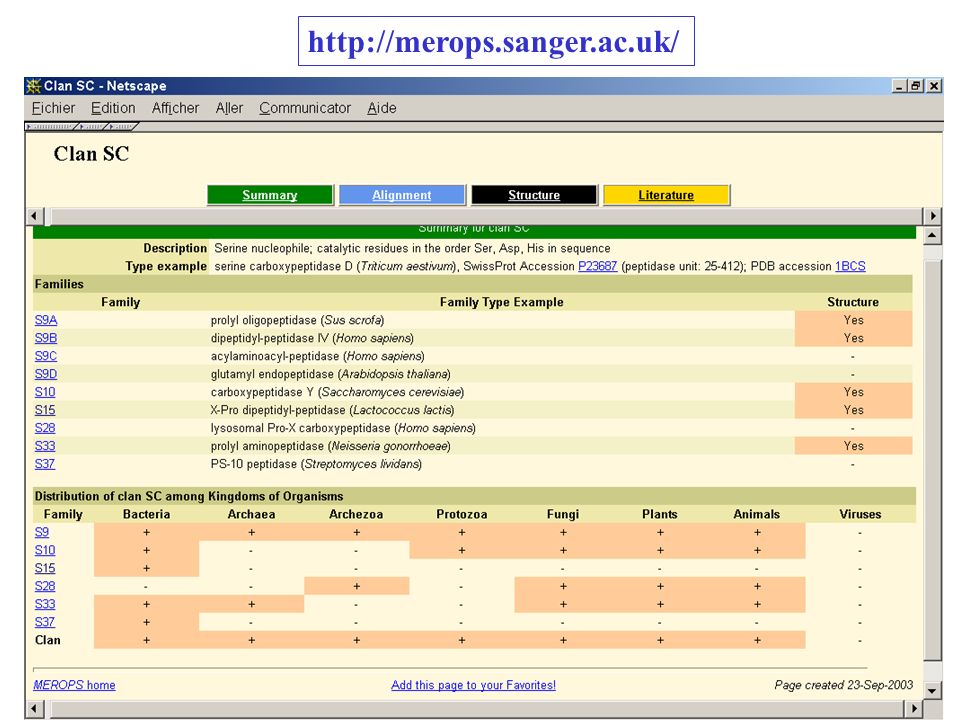
SC Clan Almost all enzymes are specialized in cleavages involving a proline residue -Oligopeptidases [endo, release peptides] -Iminopeptidases [exo, release PRO] -Carboxypeptidases [release peptides] -X-prolyl dipeptidyl aminopeptidases [exo, releases X-PRO] endo detected in the case of DDP-IV
![SC Clan Almost all enzymes are specialized in cleavages involving a proline residue -Oligopeptidases [endo, release peptides] -Iminopeptidases [exo, release PRO] -Carboxypeptidases [release peptides] -X-prolyl dipeptidyl aminopeptidases [exo, releases X-PRO] endo detected in the case of DDP-IV](http://images.slideplayer.fr/3/1288415/slides/slide_5.jpg "SC Clan Almost all enzymes are specialized in cleavages involving a proline residue -Oligopeptidases [endo, release peptides] -Iminopeptidases [exo, release PRO] -Carboxypeptidases [release peptides] -X-prolyl dipeptidyl aminopeptidases [exo, releases X-PRO] endo detected in the case of DDP-IV")
6
http://merops.sanger.ac.uk/
7
S15 family - Alignments - + legend figure 1 HERE!

8
Structure Resolution of PepX from Lactoccocus lactis 10 % PEG 4000, 150mM NaCl Ph 5.2 MES NaOH, 18°C

9
PepX prototype de la famille S15 - 4 domaines - hydrolase fold - éléments remarquables (peptide lasso, Boucle C-ter, …) helical N-terminal catalytic C-terminal lasso 85 Å25 Å Rigolet, P. et al. (2002). Structure 10, 1383-1394
. Structure 10,")
10
Enzymes families of SC Clan and related structures Catalytic domain ( hydrolase fold) in green, N-ter domain in red, C-ter domain in blue and helical domain in orange
 in green, N-ter domain in red, C-ter domain in blue and helical domain in orange")
11
Comparison of sequence and structures of SC Clan enzymes SPAP [317 residues] S33 family CBPY [416 residues] S10 family POP [710 residues] S9A family DPP-IV [726 residues] S9B family CBPY [416 residues] S10 family 228 CA (3.03 Å) 17.5 % POP [710 residues] S9A family 180 CA (3.17 Å) 15.3 % 154 CA (3.47 Å) 17.6 % DPP-IV [726 residues] S9B family 207 CA (3.11 Å) 10.8 % 189 CA (2.95 Å) 11.9 % 451 CA (3.50 Å) 19.8 % PepX [763 residues] S15 family 175 CA (3.02 Å) 16.4 % 184 CA (3.30 Å) 12.3 % 207 CA (2.63 Å) 16.8 % 201 CA (3.15 Å) 17.8 %
![Comparison of sequence and structures of SC Clan enzymes SPAP [317 residues] S33 family CBPY [416 residues] S10 family POP [710 residues] S9A family DPP-IV [726 residues] S9B family CBPY [416 residues] S10 family 228 CA (3.03 Å) 17.5 % POP [710 residues] S9A family 180 CA (3.17 Å) 15.3 % 154 CA (3.47 Å) 17.6 % DPP-IV [726 residues] S9B family 207 CA (3.11 Å) 10.8 % 189 CA (2.95 Å) 11.9 % 451 CA (3.50 Å) 19.8 % PepX [763 residues] S15 family 175 CA (3.02 Å) 16.4 % 184 CA (3.30 Å) 12.3 % 207 CA (2.63 Å) 16.8 % 201 CA (3.15 Å) 17.8 %](http://images.slideplayer.fr/3/1288415/slides/slide_11.jpg "Comparison of sequence and structures of SC Clan enzymes SPAP [317 residues] S33 family CBPY [416 residues] S10 family POP [710 residues] S9A family DPP-IV [726 residues] S9B family CBPY [416 residues] S10 family 228 CA (3.03 Å) 17.5 % POP [710 residues] S9A family 180 CA (3.17 Å) 15.3 % 154 CA (3.47 Å) 17.6 % DPP-IV [726 residues] S9B family 207 CA (3.11 Å) 10.8 % 189 CA (2.95 Å) 11.9 % 451 CA (3.50 Å) 19.8 % PepX [763 residues] S15 family 175 CA (3.02 Å) 16.4 % 184 CA (3.30 Å) 12.3 % 207 CA (2.63 Å) 16.8 % 201 CA (3.15 Å) 17.8 %")
12
Cocaine Esterase (COCE,) 1JU3, 565 residues 40% Specific to PepX between PepX and : Prolyl Iminopeptidase (XCPIP, Xant. campestris) 1JU3, 313 residues 66% Specific to PepX Prolyl Oligopeptidase (POP, porcine muscle) 1JU3, 710 residues 64% Specific to PepX Structure comparisons
 1JU3, 313 residues 66% Specific to PepX Prolyl Oligopeptidase (POP, porcine muscle) 1JU3, 710 residues 64% Specific to PepX Structure comparisons.")
13
Structure-based sequence aligment Only 4 conserved sequences can notably be distinguished between the two sequences of PepX and DPP-IV: - sequence NxxxAxxGxSYxG around the active serine ; - sequence LxxHGxxDxNVxxxxQxxxxxKAL around the active aspartic acid ; - short sequence HxxxxxS after the active histidine ; - sequence AxAxxSxWxxY before the Pos1 subsite of the X-PDAP signature.

14
PepX Dimeric structure -Important for activity -Globular shape -Involve principally N-ter and helical domains -Canal acces to catalytic residues -The two active sites are far away from each other and independently accessible to the substrate -Hydrophylic interface; labile contacts

15
Engel, M., Hoffmann, T., Wagner, L., Wermann, M., Heiser, U., Kiefersauer, R., Huber, R., Bode, W., Demuth, H.U. and Brandstetter H. (2003). Proc. Natl. Acad. Sci. USA 100, 5063-5068. DPP-IV Dimeric structure DPP-IV
. Proc. Natl. Acad. Sci. USA 100, DPP-IV Dimeric structure DPP-IV.")
16
PepX Electrostatic properties Acidic surface (also seen for other proteases of Lact. lactis ; adaptation to a particular cellular environment)
.")
17
Rasmussen HB, Branner S, Wiberg FC, Wagtmann N. (2003). Nature Struct. Biol. 10, 19-25. DPP-IV Electrostatic properties DPP-IV

18
Differences between the two enzymes -Nearly the same lenght (PepX 763 and DPP-IV 766) -Different folds (moreover 4 versus 3 domains) -Both dimer but of different quaternary organization -DPP-IV is integrated in the plasmatic membrane whereas PepX is an cytoplasmic enzyme -Substrate selection processes via an N-ter propeller domain whereas via dimeric domain in PepX.
 -Different folds (moreover 4 versus 3 domains) -Both dimer but of different quaternary organization -DPP-IV is integrated in the plasmatic membrane whereas PepX is an cytoplasmic enzyme -Substrate selection processes via an N-ter propeller domain whereas via dimeric domain in PepX.")
19
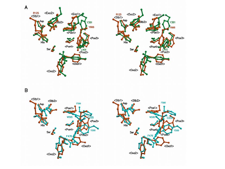
Comparison of the specificity sites of PepX and DPP IV Evolution conserved thus a particular arrangement of residues, perhaps the most efficient, ensuring XPDAP activity with a high specificity.

20
Comparison of the specificity sites of PepX and DPP IV Evolution conserved thus a particular arrangement of residues, perhaps the most efficient, ensuring XPDAP activity with a high specificity. Oxyanion hole

21
Comparison of the specificity sites of PepX and DPP IV Evolution conserved thus a particular arrangement of residues, perhaps the most efficient, ensuring XPDAP activity with a high specificity. Signature X-PDAP activity

22
Comparison of the specificity sites of PepX and DPP IV Evolution conserved thus a particular arrangement of residues, perhaps the most efficient, ensuring XPDAP activity with a high specificity. ?

24
Label PepX [family S15] DPP-IV [ family S9B] POP [family S9A] SPAP [family S33] CBPY [family S10] Catalytic triad SerS348S630S554S113S146 AspD468D708D641D268D338 HisH498H740H680H296H397 Residues implicated in positioning of the substrate proline in the active site Pos1Y380Y662W595 -- Pos2 L401Y666F476E232W312 Pos3W377W659 - L141 - Pos4I374V656V580F139L178 Residues stabilizing the binding of the substrate in the specificity pocket Stb1 - R125 R643 - Y256 Stb2N470N710R643A270I340 Oxyanion hole Oxa1Y349Y631N555W114Y147 Oxa2Y210Y547Y473 -- Residues responsible for the exopeptidase activity Exo1F393E205 --- Exo2E396E206 --- Other residues postulated to play a role in enzyme specificity OtherV471V711V644C271C341 Equivalent residues in compared enzymes of the clan SC. Structural signature of the XPDAP activity Rigolet P. et al.. (2005). FEBS J. 272; 2050-2059.
![Label PepX [family S15] DPP-IV [ family S9B] POP [family S9A] SPAP [family S33] CBPY [family S10] Catalytic triad SerS348S630S554S113S146 AspD468D708D641D268D338 HisH498H740H680H296H397 Residues implicated in positioning of the substrate proline in the active site Pos1Y380Y662W Pos2 L401Y666F476E232W312 Pos3W377W659 - L141 - Pos4I374V656V580F139L178 Residues stabilizing the binding of the substrate in the specificity pocket Stb1 - R125 R643 - Y256 Stb2N470N710R643A270I340 Oxyanion hole Oxa1Y349Y631N555W114Y147 Oxa2Y210Y547Y Residues responsible for the exopeptidase activity Exo1F393E Exo2E396E Other residues postulated to play a role in enzyme specificity OtherV471V711V644C271C341 Equivalent residues in compared enzymes of the clan SC.](http://images.slideplayer.fr/3/1288415/slides/slide_24.jpg "Structural signature of the XPDAP activity Rigolet P. et al.. (2005). FEBS J. 272;")
25
- Exploiter différences : R125 DPP-IV L401 PepX / Y666 DPPIV Recherche dinhibiteurs spécifiques -Tester inhibiteurs de DPPIV (ceux de petite taille) - Structure de PepX avec un inhibiteur de DPP-IV
 - Structure de PepX avec un inhibiteur de DPP-IV")
26
EnzymePepXDPP-IV CompoundIC 50 KIKI KIKI SI a valine-pyrrolidide 30 M9.3 M 4 M b 2 M c 7.5 diprotin A 260 M71.5 M 8 M d 4.6 M e 32.5 diprotin B 600 M 118.45 M Table 2: Inhibition experiments realized with PepX from Lactococcus lactis. Recherche dinhibiteurs spécifiques

27
Valine-Pyrrolidide Inhibition compétitive IC 50 = 30 M K I = 9 M Rigolet P., Xi, X.G., Rety, S. and Chich, J.F. (2005). FEBS J. 272; 2050-2059. K I = 9 M Recherche dinhibiteurs spécifiques
. FEBS J. 272; K I = 9 M Recherche dinhibiteurs spécifiques.")
28
Table 3: Docking simulations of valine-pyrrolidide in the X-PDAP enzymes. Recherche dinhibiteurs spécifiques EnzymePepXDPP-IV CompoundIC 50 KIKI KIKI SI a valine-pyrrolidide 30 M9.3 M 4 M b 2 M c 7.5 diprotin A 260 M71.5 M 8 M d 4.6 M e 32.5 diprotin B 600 M 118.45 M Table 2: Inhibition experiments realized with PepX from Lactococcus lactis.

29
Recherche dinhibiteurs spécifiques Nouveaux dérivés partant de la valine-pyrrolidide … … Nous les avons synthétisés …. … Nous les testons actuellement ….

30
Drug design: autres stratégies -boucles C-TER - interface de dimérisation - Mutagenèse dirigée : PHE 80

31
Quelle est la fonction des domaines Nter et Cter? -Quapportent-ils vraiment à la catalyse ? -Rôle présenté chez DPPIV -Pas de réelle interprétation ni chez aAeH ni chez Coce -Rôle supposé chez PepX, qui possède à la fois un Nter et un Cter -Siège d'une deuxième fonction ?

32
P. RigoletINRA – LURE – ENS Cachan J. F. ChichINRA M. M. DelageINRA I. MechinEMBL Contributions Collaboration avec CHIMISTES de lENS-Cachan Equipe de Joane XIE Synthèse organique d'inhibiteurs potentiels

Présentations similaires