Télécharger la présentation
La présentation est en train de télécharger. S'il vous plaît, attendez

1
M.A. Mahé, A. Lisbona, E. Kantor, P. Giraud
Tomothérapie M.A. Mahé, A. Lisbona, E. Kantor, P. Giraud Nantes, Bordeaux, Paris GRO, Dinan le 23/01/2010

2
Plan Principes Expérience française préliminaire
Tomothérapie versus Arcthérapie dynamique

3
Implementation of Tomotherapy in France
Lille 2005: Request for proposals: innovative techniques in radiotherapy 2007: Paris Nantes Bordeaux 2008: Strasbourg 2009: Toulouse Lille (x2) Rouen Caen Reims Strasbourg Nancy Brest Rennes Ile de France Dijon Besançon Nantes Poitiers Lyon Limoges Grenoble St Etienne – Ferrand Bordeaux Nice 1 Montpellier Toulouse Marseille
 Rouen. Caen. Reims. Strasbourg. Nancy. Brest. Rennes. Ile de France. Dijon. Besançon. Nantes. Poitiers. Lyon. Limoges. Grenoble. St Etienne. – Ferrand. Bordeaux. Nice. 1. Montpellier. Toulouse. Marseille.")
4
TomoTherapy HIART 85cm same as ct simulators, patients up to 200 kg
We can probably do patients that other manufacurors can not do (normally don’t do that) Imaging FOV 40x160, (treating bigger field even) Not a lot of controls, no pendants from ceiling, no couch controls, most controls done by computer others from touch screens on gantry
 Imaging FOV 40x160, (treating bigger field even) Not a lot of controls, no pendants from ceiling, no couch controls, most controls done by computer others from touch screens on gantry.")
5
Radiothérapie guidée par l’image (IGRT)
Le principe physique est celui du scanner hélicoïdal en imagerie. Le tube à rayons X de haute énergie tourne autour du patient pendant que le lit de traitement se déplace dans le tunnel. Ce équipement permet également de contrôler le positionnement du patient, à chaque séance, par une imagerie du type de celle fournie par le scanner en radiologie. 85 cm

6
Le faisceau est mis en forme à l'aide d'une fente permettant sa collimation et la définition d'un champ d’irradiation de 40 cm de longueur par 1, 2,5 ou 5 cm de largeur couvrant ainsi une «tranche»

7
Radiothérapie conformationnelle avec modulation d’intensité (RCMI)
Chaque secteur de la «tranche» du faisceau est modulé à volonté au cours de la rotation par un système de collimation additionnelle composé de 64 lames de 0,61 cm opposées et fonctionnant chacune sur un mode binaire ouvert ou fermé (arrêtant ou laissant passer les rayonnements).
.")
8
Traitement Helicoidal jusqu’à 160 cm de longueur
Epaisseur de coupe Serial tomotherapy, nomos peackock system. Not to explane always Pitch 0,1 O,5 and 0,9 Principaux Pitches 0,2 à 0,5 Traitement 1 à 2 Imagerie Pitch = Déplacement parcourue par la table et par rotation de 360° Epaisseur de coupe T-MKT-AP0068

9
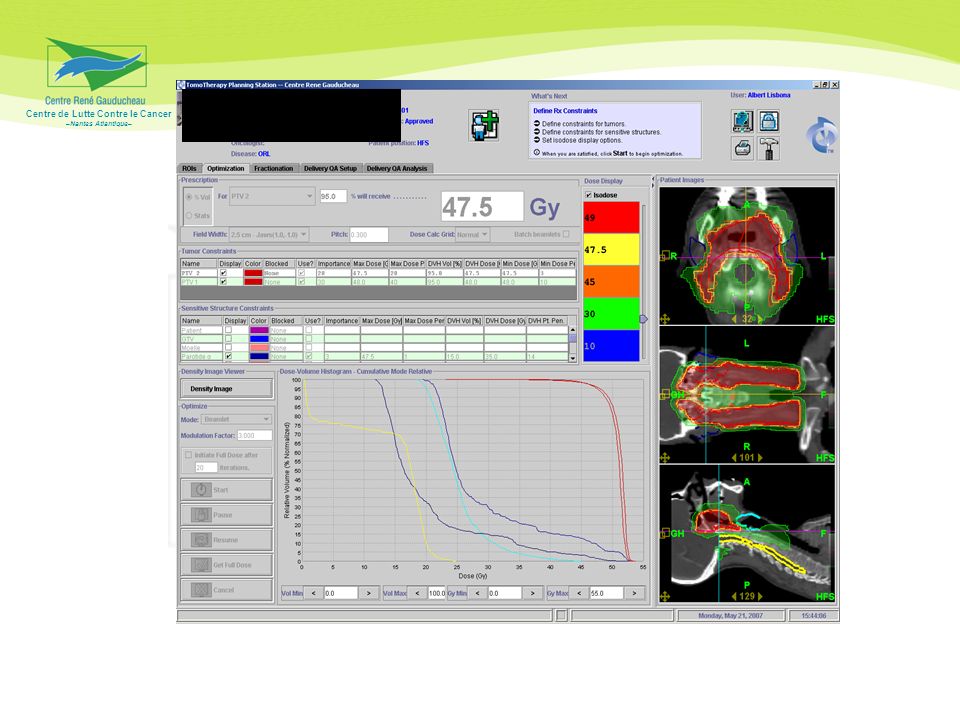
Expérience française 01/2007 – 06/2009 606 points analysables
Suivi moyen 7-9 mois (1-29 mois)
")
10
Protocole d’évaluation
INTRODUCTION 4 SYNOPSIS TOMOTHÉRAPIE PAR ÉTUDE ET LOCALISATION 1. SARCOMES 1.1. SARCOMES DES MEMBRES ET PAROI DE L’ADULTE 5 1.2. SARCOMES RÉTRO–PÉRITONÉAUX DE L’ADULTE 9 1.3. TUMEURS AXIALES, PARA–AXIALES ET PELVIENNES DE L’OS ET DES PARTIES MOLLES DE L’ADULTE 13 2. MÉNINGIOMES INTRACRÂNIENS DE GRADE 1 et AUTRES TUMEURS CÉRÉBRALES NON GLIALES 17 3. PROSTATE 22 4. CANCERS ORL 25 5. ICT 30 6. SEIN 31 7. TUMEURS THORACIQUES 7.1. CARCINOMES BRONCHIQUES NON À PETITES CELLULES 36 7.2. MÉSOTHÉLIOMES 41 8. MÉTASTASES OSSEUSES 47 9. IRRADIATION CRÂNIO–SPINALE DE L’ADULTE 49 10. CANAL ANAL 53 ANNEXES 57

11
Total dose Number of fractions Dose per fraction Anal canal 59.4 Gy 33 1.8 Gy Bone metastasis 8 Gy 1 Breast Gy 25-29 1.8-2 Gy Cranio-spinal irradiation 36 Gy (Optional posterior fossa boost of 18 Gy) 18 (+9) 2 Gy Head and Neck Gy 28-35 2-2.3 Gy Lung tumors (Non small cell) 68 Gy 34 Meningioma and other CNS tumors (except for glial tumors) 54 Gy 30 Mesothelioma 50-54 Gy 25-30 Prostate 70-80 Gy 35-40 Sarcoma, axial 45-65 Gy 25-36 Sarcoma, limb and chest wall Sarcoma, retroperitoneal
 18 (+9) 2 Gy. Head and Neck Gy Gy. Lung tumors (Non small cell) 68 Gy. 34. Meningioma and other CNS tumors (except for glial tumors) 54 Gy. 30. Mesothelioma Gy Prostate Gy Sarcoma, axial Gy Sarcoma, limb and chest wall. Sarcoma, retroperitoneal.")
12
Middle and Internal ear 54 Gy Retina Lens 8 Gy Peripheral nerve plexus
Dose constraints Nervous system: Brain 1/3 < 54 Gy Brainstem 6 ml < 54 Gy Pituitary 50 Gy Spinal cord 45 Gy Middle and Internal ear 54 Gy Retina Lens 8 Gy Peripheral nerve plexus 54 Gy. Head and Neck: Parotid 26 Gy Temporomandibular joint 60 Gy Mandible 70 Gy Thorax: Lung V20 Gy < 25% Esophagus V50 Gy < 35% Heart V40 Gy < 30% Abdomen: Liver V30 Gy < 40% Kidney One kidney < 12 Gy Small bowel and Colon Max dose: 54 Gy; V45 Gy < 1/3; V20 Gy < 1/2 Pelvis: Rectum Max dose: 76 Gy; 1/4 > 72 Gy Bladder Max dose: 80 Gy; 1/2 > 70 Gy Femoral heads 5% > 55 Gy Gonads 2 Gy

13
Number of patients Sexe Male 346 Female 260 Age <18 22 18-30 38 31-50 130 51-70 320 >71 96 Regional site of disease CNS 39 Head and Neck 196 Thorax and Chest wall 79 Abdomen 45 Pelvis 123 Bone and Soft tissue 109 Multiple sites 15 Treatment intent Curative 531 Palliative 75 Surgery Pre-operative RT 33 Post-operative RT 228 No surgery 345 Chemotherapy Adjuvant CT alone 128 Concomitant CT alone 102 Neoadjuvant and Concomitant CT 73 No CT 303 Re-irradiation Yes 78 No 528

14
Acute toxicity Chronic toxicity grade 3 grade 4 grade 5 CNS 1 Head and Neck 38 2 Thorax and Chest wall Abdomen 4 Pelvis 14 3 Bone and Soft tissue Multiple sites 5 Total 66 8

15
Tomothérapie - ORL Données de la littérature Van Vulpen et al
Tomothérapie - ORL Données de la littérature Van Vulpen et al. Int J Radiat Oncol Biol Phys 2005; 62: 5 oropharynx Comparaison dosimétrique : RCMI step and shout – Tomo 69 Gy Dose moyenne parotides : Tomo 24 Gy RCMI 30 Gy

16
Tomothérapie - ORL Données de la littérature Fiorino et al
Tomothérapie - ORL Données de la littérature Fiorino et al. Radiother Oncol 2006; 78: 3 oropharynx, 1 hypopharynx, 1 larynx Comparaison dosimétrique : RCMI dynamique – Tomo PTV1 54 Gy Tomo RCMI PTV1 V95% Parotides dose moy Moelle (dose max) 96,2 % 20,8 Gy 24,5 Gy 89,6 % 26,3 Gy 31,6 Gy
 96,2 % 20,8 Gy. 24,5 Gy. 89,6 % 26,3 Gy. 31,6 Gy.")
19
Toxicité cutanée

21
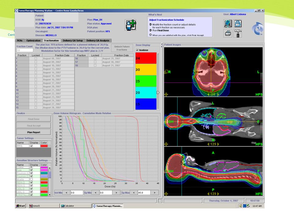
Médulloblastoma

22
22

23
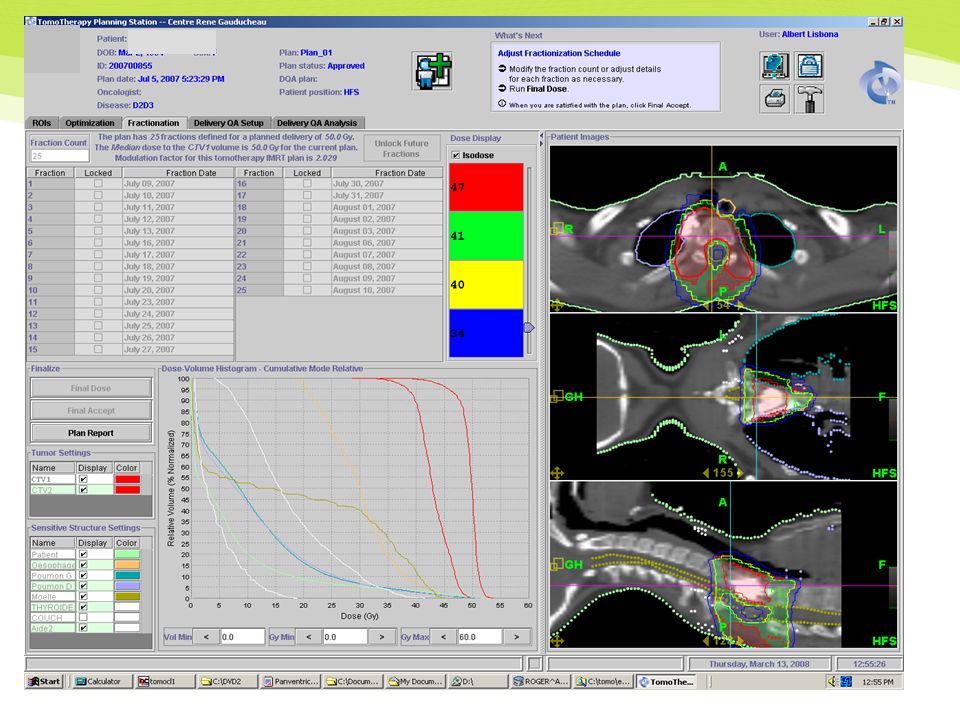
Craniospinal irradiation
DOSIMETRIC DATA Organs Mean dose CS-TOMO Mean dose CS-RT Mean dose ratio PTV brain 35.8 37.3 1.04 PTV spine 35.9 38.1 1.06 Parotid 10.4 20.1 1.93 Thyroid 9.7 23.2 2.39 Esophagus 9.8 32.2 3.29 Heart 6.4 14.6 2.27 Bowel 9.0 11.6 1.29

25
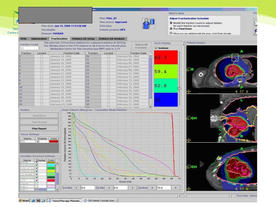
Mesothelioma Post operative hemithoracic irradiation
PTV = hemithorax + scar: 50 Gy +/- residual tumor: 4 Gy OAR: heart kidneys liver lung+++ V20 < 7% (Rice IJROBP 2007; 69: )
")
26
Mesothelioma pN0

27
Mesothelioma pN2

28
Anal cancer

29
Endometrial cancer

35
Whole ventricular irradiation Pediatric germ cell tumors Supiot et al, ASTRO 2008
Objective: Dosimetric comparison 2 fields conformal RT 5 fields conformal RT 5 fields IMRT 24 Gy/12 f (excluding tumor boost) Helical IMRT Methods 6 patients volume PTV = CTV ventricles + 5mm + CTV tumor + 2mm
 Helical IMRT. Methods. 6 patients. volume. PTV = CTV ventricles + 5mm. + CTV tumor. + 2mm.")
36
Whole ventricular irradiation
conformal 2 fields conformal 5 fields IMRT 5 fields Helical IMRT p (two-ways anova) chiasm Mean dose 23.0 Gy 23.1 22.1 17.8 0.025 Temporal lobes Mean dose 21.5 Gy 19.6 Gy 17.9 Gy 16.5 Gy <0.0001 pituitary Mean Dose 21.9 Gy 22.6 Gy 21.1 Gy 14.3 Gy 0.0157 Brain stem Mean Dose 19.6 Gy 20.2 Gy 21.9 Gy 17.6 Gy 0.15
 chiasm. Mean dose Gy Temporal lobes. Mean dose Gy Gy Gy Gy. < pituitary. Mean Dose Gy Gy Gy Gy Brain stem. Mean Dose Gy Gy Gy Gy")
37
Conformal RT : 2 fields Exemple de dosi par 2 fx : un tunnel

38
Helical IMRT Bonne couverture homogène du volume cible

39
Low doses and IMRT: V2Gy Whole ventricular irradiation: 5 children
Conformal 2 fields Conformal 5 fields IMRT 5 fields Helical IMRT p Face 2Gy 31% 90% 91% 100% <0.0001

40
TBI Questions Organes à protéger ? Débit de dose : Tomo = 6-8 Gy / min
Classique = cGy /mn

41
Irradiation médullaire totale (IMT)
Wong et al, biology of blood and marrow transplantation, 12: , 2006 Étude dosimétrique Organes IMT 10 Gy IMT 20 Gy ICT conventionnelle Tomo 12 Gy Poumons 4,3 6,8 8,8 Foie 6 8,7 12,4 Reins 5,6 12,2 Cœur 6,2 6,4 12,1 Thyroïde 3,7 4,9 Cristallins 1,5 1,9 11,3 Parotides 3,9 4,8 11,8

42
IMT MM stades I – III répondeurs ou stables après chimio
Wong JY et al, IJROBP; 73: , 2009 MM stades I – III répondeurs ou stables après chimio Melphalan - CSP IMT – CSP : Phase I 13 patients : 10, 12, 14, 16 Gy (2 Gy x 2/j) Toxicité grade 1 – 2 : vomissement, mucite, fatigue, diarrhée Toxicité grade 3 – 4 : 0
 Toxicité grade 1 – 2 : vomissement, mucite, fatigue, diarrhée. Toxicité grade 3 – 4 : 0.")
43
Fig. 1. Sample treatment plans for the same patient with either (a) helical tomotherapy or (b) topotherapy (static fields). The following isodose lines are displayed: 20 Gy, 30 Gy, 40 Gy, 50.4 Gy (prescription dose) and 52.9 Gy (105% of prescription dose). The dose prescription was to the V95%.
 helical tomotherapy or (b) topotherapy (static fields). The following isodose lines are displayed: 20 Gy, 30 Gy, 40 Gy, 50.4 Gy (prescription dose) and 52.9 Gy (105% of prescription dose). The dose prescription was to the V95%..")
44
Fig. 2. Dose–volume histograms for the same patient treated with either (a) helical tomotherapy or (b) topotherapy (static fields).
 helical tomotherapy or (b) topotherapy (static fields)..")
45
Temps d’irradiation Loc. Moyenne (mn) Extrêmes (mn) ORL 7 3.6 - 11.5
SNC 4.3 Thorax Abdomen 9.7 Pelvis 5.7 Os-tissus mous 6.7 15 – 23 pts/J – 10h/J

46
Temps d’irradiation Bauman Médiane : 6mn (2-68) N : 60 Sterzing
Moyenne : 10.7mn (6.8 – 46.4mn) N: 150
 N: 150.")
47
Thomothérapie versus Arcthérapie dynamique
Evaluation médico-économique Tomo vs Rapid Arc vs VMAT Objectif principal Différentiel de coûts Objectifs secondaires Impact de l’apprentissage Toxicité Contrôle local, SSR, SG Distribution de dose Etude ORL: début 2010 Etude pelvis : (prostate, col utérin, canal anal avec RT gg) : mi 2010
 : mi")
48
Conclusion Tomo : grands volumes, volumes complexes, ré-irradation, 2ème K en territoire irradié Toxicité aigue et semi-tardive acceptable Tomo vs Arcthérapie dynamique : attendre résultats EME

Présentations similaires


.>")
 Mitchell 5 tables (25 étuis) Tables: 5 Rondes: 5 de 5 étuis Étuis au jeu: 25 Fantôme: 5 N-S (ou E-O) Select movement:>")

