Télécharger la présentation
La présentation est en train de télécharger. S'il vous plaît, attendez

1
Jean-François TIMSIT CHU Grenoble UJF/Inserm U 823
Conduite à tenir devant une suspicion d’infection liée aux cathéters en réanimation Jean-François TIMSIT CHU Grenoble UJF/Inserm U 823 St Etienne – Juin 2009

2
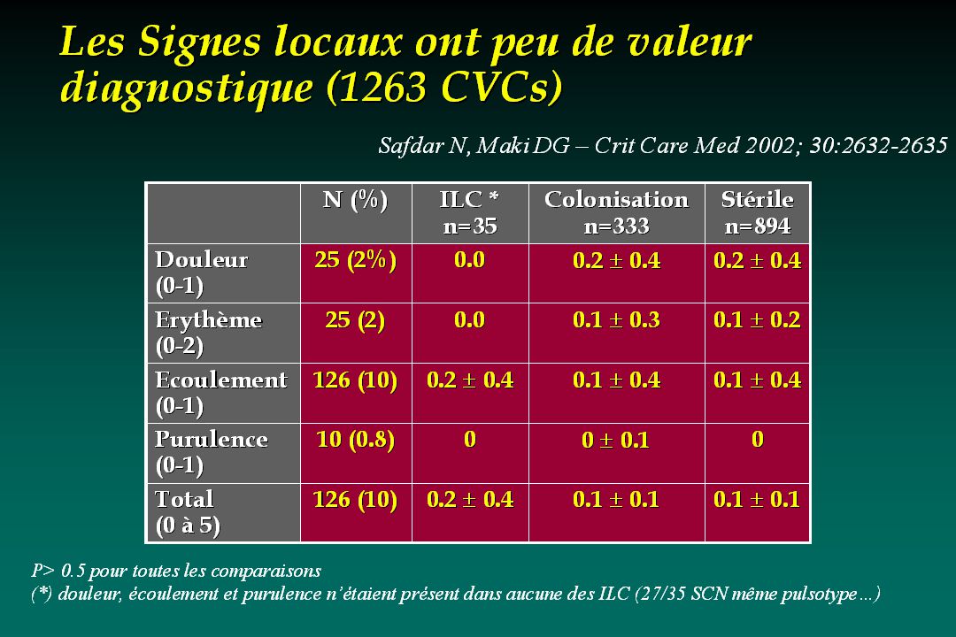
ILC : Le traitement depend de
sévérité du sepsis maladies sous-jacente (immunodépression, prothèses). Micro-organismes identifiés ou suspectés HC positives ou négatives Utilité et facilité de l’abord veineux central Faible niveau de preuve 1
. Micro-organismes identifiés ou suspectés. HC positives ou négatives. Utilité et facilité de l’abord veineux central. Faible niveau de preuve. 1.")
3
En cas de sepsis grave le cathéter DOIT être enlevé
Deux constraintes : Eviter l’ablation inutile des CVCs (75% cases) et le risque associé de complications mécaniques Sauver les malades et éviter que l’infection se complique En cas de sepsis grave le cathéter DOIT être enlevé 8
 et le risque associé de complications mécaniques. Sauver les malades et éviter que l’infection se complique. En cas de sepsis grave le cathéter DOIT être enlevé. 8.")
4
2 situations Fièvre sans signes de sepsis sévère en réanimation
Sepsis sévère de cause inconnu Ablation du CVC (ou échange sur guide?) Quels antibiotiques? Comment dépister les complications et les traiter? Fièvre sans signes de sepsis sévère en réanimation Hémoculture positive Est il possible de conserver le cathéter sans risques?
 Quels antibiotiques Comment dépister les complications et les traiter Fièvre sans signes de sepsis sévère en réanimation. Hémoculture positive. Est il possible de conserver le cathéter sans risques")
5
Le cathéter? Ablation du cathéter Est associée à un plus grand nombre de guérison et une amélioration du pronostic 2. Diagnostic cathéter en place 3. Echange sur guide (GWX)
")
6
Schneegurt, MA. Wichita St. University, Microbiology 103.
Biofilm formation Schneegurt, MA. Wichita St. University, Microbiology 103.

7
Why form a bioflim? Jefferson KK. FEMS. 2004;236:

8
Susceptibility of biofilm organisms
Antibiotic MIC or MBC (mcg/mL) Effective [ ] vs. biofilm (mcg/mL) S. aureus (NCTC ) Vancomycin 2 (MBC) 20 P. aeruginosa (ATCC 27853) Imipenem 1 (MIC) >1,024ª E. coli (ATCC 25922) Ampicillin 2 (MIC) 512ª P. pseudomallei Ceftazidime 8 (MBC) 800 S. sanguis Doxycycline 0.063 (MIC) 3.15 ª Minimal biofilm eradication Adapted from Donlan RM, et al. Clin Microbiol Rev. 2002;15:
 Effective [ ] vs. biofilm (mcg/mL) S. aureus. (NCTC ) Vancomycin. 2 (MBC) 20. P. aeruginosa (ATCC 27853) Imipenem. 1 (MIC) >1,024ª. E. coli. (ATCC 25922) Ampicillin. 2 (MIC) 512ª. P. pseudomallei. Ceftazidime. 8 (MBC) 800. S. sanguis. Doxycycline (MIC) ª Minimal biofilm eradication. Adapted from Donlan RM, et al. Clin Microbiol Rev. 2002;15:")
9
CVC maintenance is always risky…
1- Bacterias with slime production have an increased MICs and MBCs to ABx 2- The Biofilm increases the resistance of bacteria to ABt SCN culture Here is the MIC s of a slime producer Sraf epidermidis. For example the MIC of gentamycin was increased by more than a hundered fold in presence of slime. The MBCs of attached bacteria are also increased by a more than 128 fold. Finaly CVC maintenance is always associated with an increased risk of failure in case of catheter-related sepsis . However in three forth of the cases, catheters are removed but innocent!!! CVC maintenance is always risky…

10
Catheter removal and duration of candidemia
Rex et al -Decrease of the duration of the candidemia New site 5.6 days vs Other 2.6 days - Bias: APACHE II 14.5 vs 16.9 p=0.03 Other catheter: 1.2 vs 1.8,p<0.001 - GWX: j Post hoc analysis of a randomized study comparing fluconazole and amphotericine B Catheter removal should be prefered

11
Management of CVCs in patients with cancer and candidemia
Management of CVCs in patients with cancer and candidemia Raad I et al – Clin Infect Dis 2004; 38:1119 : 404 episodes of candidemia (50% ICU) with 1 CVCs for more than 1 days 3 categories Primary candidemia : 241 (60%) Secondary candidemia: 52 (13%) CVC related candidemia : 111 (27%) + tip cult (66) or quantitative BC > 5:1 (45) % Management of Central Venous Catheters in Patients with Cancer and Candidemia Issam Raad,1 Hend Hanna,1 Maha Boktour,1 Essam Girgawy,1 Hadi Danawi,3 Masoud Mardani,1 Dimitrios Kontoyiannis,1 Rabih Darouiche,2 Ray Hachem,1 and Gerald P. Bodey1 1Department of Infectious Diseases, Infection Control and Employee Health, University of Texas M.D. Anderson Cancer Center, 2Baylor College of Medicine, and 3University of Texas School of Public Health, Houston To determine the need and appropriate timing of catheter removal in patients with candidemia, the records for 404 patients with cancer and central venous catheters (CVCs) who developed candidemia during the period of were retrospectively reviewed. Of the total 404 cases of candidemia, 241 (60%) were due to a primary source, 111 (27%) were catheter related, and 52 (13%) were secondary cases of candidemia caused by a source other than the catheter. Multivariate analysis showed that catheter removal 72 h after onset improved response to antifungal therapy exclusively in patients with catheter-related candidemia (P = .04). Clinical characteristics that suggested a noncatheter source for the candidemia were disseminated infection (P < .01), previous chemotherapy (P < .01), previous corticosteroid therapy (P = .02), and poor response to antifungal therapy (P < .03). CVC removal 72 h after onset should be considered in patients with suspected catheter-related candidemia who have no evidence of dissemination, recent corticosteroid therapy, or chemotherapy.
 with 1 CVCs for more than 1 days. 3 categories. Primary candidemia : 241 (60%) Secondary candidemia: 52 (13%) CVC related candidemia : 111 (27%) + tip cult (66) or quantitative BC > 5:1 (45) % Management of Central Venous Catheters in Patients with Cancer and Candidemia. Issam Raad,1 Hend Hanna,1 Maha Boktour,1 Essam Girgawy,1 Hadi Danawi,3 Masoud Mardani,1 Dimitrios Kontoyiannis,1 Rabih Darouiche,2 Ray Hachem,1 and Gerald P. Bodey1. 1Department of Infectious Diseases, Infection Control and Employee Health, University of Texas M.D. Anderson Cancer Center, 2Baylor College of Medicine, and 3University of Texas School of Public Health, Houston. To determine the need and appropriate timing of catheter removal in patients with candidemia, the records for 404 patients with cancer and central venous catheters (CVCs) who developed candidemia during the period of were retrospectively reviewed. Of the total 404 cases of candidemia, 241 (60%) were due to a primary source, 111 (27%) were catheter related, and 52 (13%) were secondary cases of candidemia caused by a source other than the catheter. Multivariate analysis showed that catheter removal 72 h after onset improved response to antifungal therapy exclusively in patients with catheter-related candidemia (P = .04). Clinical characteristics that suggested a noncatheter source for the candidemia were disseminated infection (P < .01), previous chemotherapy (P < .01), previous corticosteroid therapy (P = .02), and poor response to antifungal therapy (P < .03). CVC removal 72 h after onset should be considered in patients with suspected catheter-related candidemia who have no evidence of dissemination, recent corticosteroid therapy, or chemotherapy.")
12
Outcome of candidemia: time of catheter removal after the first positive culture Raad I et al – Clin Infect Dis 2004; 38:1119 Management of Central Venous Catheters in Patients with Cancer and Candidemia Issam Raad,1 Hend Hanna,1 Maha Boktour,1 Essam Girgawy,1 Hadi Danawi,3 Masoud Mardani,1 Dimitrios Kontoyiannis,1 Rabih Darouiche,2 Ray Hachem,1 and Gerald P. Bodey1 1Department of Infectious Diseases, Infection Control and Employee Health, University of Texas M.D. Anderson Cancer Center, 2Baylor College of Medicine, and 3University of Texas School of Public Health, Houston To determine the need and appropriate timing of catheter removal in patients with candidemia, the records for 404 patients with cancer and central venous catheters (CVCs) who developed candidemia during the period of were retrospectively reviewed. Of the total 404 cases of candidemia, 241 (60%) were due to a primary source, 111 (27%) were catheter related, and 52 (13%) were secondary cases of candidemia caused by a source other than the catheter. Multivariate analysis showed that catheter removal 72 h after onset improved response to antifungal therapy exclusively in patients with catheter-related candidemia (P = .04). Clinical characteristics that suggested a noncatheter source for the candidemia were disseminated infection (P < .01), previous chemotherapy (P < .01), previous corticosteroid therapy (P = .02), and poor response to antifungal therapy (P < .03). CVC removal 72 h after onset should be considered in patients with suspected catheter-related candidemia who have no evidence of dissemination, recent corticosteroid therapy, or chemotherapy.
 who developed candidemia during the period of were retrospectively reviewed. Of the total 404 cases of candidemia, 241 (60%) were due to a primary source, 111 (27%) were catheter related, and 52 (13%) were secondary cases of candidemia caused by a source other than the catheter. Multivariate analysis showed that catheter removal 72 h after onset improved response to antifungal therapy exclusively in patients with catheter-related candidemia (P = .04). Clinical characteristics that suggested a noncatheter source for the candidemia were disseminated infection (P < .01), previous chemotherapy (P < .01), previous corticosteroid therapy (P = .02), and poor response to antifungal therapy (P < .03). CVC removal 72 h after onset should be considered in patients with suspected catheter-related candidemia who have no evidence of dissemination, recent corticosteroid therapy, or chemotherapy.")
13
Predictors of failure to respond to antifungal therapy
Predictors of failure to respond to antifungal therapy Raad I et al – Clin Infect Dis 2004; 38:1119 Management of Central Venous Catheters in Patients with Cancer and Candidemia Issam Raad,1 Hend Hanna,1 Maha Boktour,1 Essam Girgawy,1 Hadi Danawi,3 Masoud Mardani,1 Dimitrios Kontoyiannis,1 Rabih Darouiche,2 Ray Hachem,1 and Gerald P. Bodey1 1Department of Infectious Diseases, Infection Control and Employee Health, University of Texas M.D. Anderson Cancer Center, 2Baylor College of Medicine, and 3University of Texas School of Public Health, Houston To determine the need and appropriate timing of catheter removal in patients with candidemia, the records for 404 patients with cancer and central venous catheters (CVCs) who developed candidemia during the period of were retrospectively reviewed. Of the total 404 cases of candidemia, 241 (60%) were due to a primary source, 111 (27%) were catheter related, and 52 (13%) were secondary cases of candidemia caused by a source other than the catheter. Multivariate analysis showed that catheter removal 72 h after onset improved response to antifungal therapy exclusively in patients with catheter-related candidemia (P = .04). Clinical characteristics that suggested a noncatheter source for the candidemia were disseminated infection (P < .01), previous chemotherapy (P < .01), previous corticosteroid therapy (P = .02), and poor response to antifungal therapy (P < .03). CVC removal 72 h after onset should be considered in patients with suspected catheter-related candidemia who have no evidence of dissemination, recent corticosteroid therapy, or chemotherapy.
 who developed candidemia during the period of were retrospectively reviewed. Of the total 404 cases of candidemia, 241 (60%) were due to a primary source, 111 (27%) were catheter related, and 52 (13%) were secondary cases of candidemia caused by a source other than the catheter. Multivariate analysis showed that catheter removal 72 h after onset improved response to antifungal therapy exclusively in patients with catheter-related candidemia (P = .04). Clinical characteristics that suggested a noncatheter source for the candidemia were disseminated infection (P < .01), previous chemotherapy (P < .01), previous corticosteroid therapy (P = .02), and poor response to antifungal therapy (P < .03). CVC removal 72 h after onset should be considered in patients with suspected catheter-related candidemia who have no evidence of dissemination, recent corticosteroid therapy, or chemotherapy.")
14
Is candidemia catheter-related
Is candidemia catheter-related? Raad I et al – Clin Infect Dis 2004; 38:1119 111 catheter-related candidemia and 52 secondary candidemia No corticosteroids within 1 month: OR 3.5 ( ), p=0.02 No chemotherapy within 1 month: OR 4.3 ( ), p<0.01 Non disseminated infection * OR 9.7 ( ), p<0.01 Good response to antifungal therapy** OR 2.9 ( ), p=0.03 Management of Central Venous Catheters in Patients with Cancer and Candidemia Issam Raad,1 Hend Hanna,1 Maha Boktour,1 Essam Girgawy,1 Hadi Danawi,3 Masoud Mardani,1 Dimitrios Kontoyiannis,1 Rabih Darouiche,2 Ray Hachem,1 and Gerald P. Bodey1 1Department of Infectious Diseases, Infection Control and Employee Health, University of Texas M.D. Anderson Cancer Center, 2Baylor College of Medicine, and 3University of Texas School of Public Health, Houston To determine the need and appropriate timing of catheter removal in patients with candidemia, the records for 404 patients with cancer and central venous catheters (CVCs) who developed candidemia during the period of were retrospectively reviewed. Of the total 404 cases of candidemia, 241 (60%) were due to a primary source, 111 (27%) were catheter related, and 52 (13%) were secondary cases of candidemia caused by a source other than the catheter. Multivariate analysis showed that catheter removal 72 h after onset improved response to antifungal therapy exclusively in patients with catheter-related candidemia (P = .04). Clinical characteristics that suggested a noncatheter source for the candidemia were disseminated infection (P < .01), previous chemotherapy (P < .01), previous corticosteroid therapy (P = .02), and poor response to antifungal therapy (P < .03). CVC removal 72 h after onset should be considered in patients with suspected catheter-related candidemia who have no evidence of dissemination, recent corticosteroid therapy, or chemotherapy. (*) Dissemination to non contiguous sites (**)Resolution of fever and chills, BC neg.
, p=0.02. No chemotherapy within 1 month: OR 4.3 ( ), p<0.01. Non disseminated infection * OR 9.7 ( ), p<0.01. Good response to antifungal therapy** OR 2.9 ( ), p=0.03. Management of Central Venous Catheters in Patients with Cancer and Candidemia. Issam Raad,1 Hend Hanna,1 Maha Boktour,1 Essam Girgawy,1 Hadi Danawi,3 Masoud Mardani,1 Dimitrios Kontoyiannis,1 Rabih Darouiche,2 Ray Hachem,1 and Gerald P. Bodey1. 1Department of Infectious Diseases, Infection Control and Employee Health, University of Texas M.D. Anderson Cancer Center, 2Baylor College of Medicine, and 3University of Texas School of Public Health, Houston. To determine the need and appropriate timing of catheter removal in patients with candidemia, the records for 404 patients with cancer and central venous catheters (CVCs) who developed candidemia during the period of were retrospectively reviewed. Of the total 404 cases of candidemia, 241 (60%) were due to a primary source, 111 (27%) were catheter related, and 52 (13%) were secondary cases of candidemia caused by a source other than the catheter. Multivariate analysis showed that catheter removal 72 h after onset improved response to antifungal therapy exclusively in patients with catheter-related candidemia (P = .04). Clinical characteristics that suggested a noncatheter source for the candidemia were disseminated infection (P < .01), previous chemotherapy (P < .01), previous corticosteroid therapy (P = .02), and poor response to antifungal therapy (P < .03). CVC removal 72 h after onset should be considered in patients with suspected catheter-related candidemia who have no evidence of dissemination, recent corticosteroid therapy, or chemotherapy. (*) Dissemination to non contiguous sites. (**)Resolution of fever and chills, BC neg.")
15
Proposed algorythm for candidemia
Proposed algorythm for candidemia Raad I et al – Clin Infect Dis 2004; 38:1119 Management of Central Venous Catheters in Patients with Cancer and Candidemia Issam Raad,1 Hend Hanna,1 Maha Boktour,1 Essam Girgawy,1 Hadi Danawi,3 Masoud Mardani,1 Dimitrios Kontoyiannis,1 Rabih Darouiche,2 Ray Hachem,1 and Gerald P. Bodey1 1Department of Infectious Diseases, Infection Control and Employee Health, University of Texas M.D. Anderson Cancer Center, 2Baylor College of Medicine, and 3University of Texas School of Public Health, Houston To determine the need and appropriate timing of catheter removal in patients with candidemia, the records for 404 patients with cancer and central venous catheters (CVCs) who developed candidemia during the period of were retrospectively reviewed. Of the total 404 cases of candidemia, 241 (60%) were due to a primary source, 111 (27%) were catheter related, and 52 (13%) were secondary cases of candidemia caused by a source other than the catheter. Multivariate analysis showed that catheter removal 72 h after onset improved response to antifungal therapy exclusively in patients with catheter-related candidemia (P = .04). Clinical characteristics that suggested a noncatheter source for the candidemia were disseminated infection (P < .01), previous chemotherapy (P < .01), previous corticosteroid therapy (P = .02), and poor response to antifungal therapy (P < .03). CVC removal 72 h after onset should be considered in patients with suspected catheter-related candidemia who have no evidence of dissemination, recent corticosteroid therapy, or chemotherapy.
 who developed candidemia during the period of were retrospectively reviewed. Of the total 404 cases of candidemia, 241 (60%) were due to a primary source, 111 (27%) were catheter related, and 52 (13%) were secondary cases of candidemia caused by a source other than the catheter. Multivariate analysis showed that catheter removal 72 h after onset improved response to antifungal therapy exclusively in patients with catheter-related candidemia (P = .04). Clinical characteristics that suggested a noncatheter source for the candidemia were disseminated infection (P < .01), previous chemotherapy (P < .01), previous corticosteroid therapy (P = .02), and poor response to antifungal therapy (P < .03). CVC removal 72 h after onset should be considered in patients with suspected catheter-related candidemia who have no evidence of dissemination, recent corticosteroid therapy, or chemotherapy.")
16
Jensen et coll Arch Intern Med 2002;162:25-32

17
Absence of CVC removal is always risky…
Candida sp: 427 CRB Death 41% vs 21 % N’guyen et al - Arch Intern Med 1995;155:2429 S. aureus: 50 CRB (retrospective) Persistent BC:11 vs 56% (p=0.01), Deaths: 5 vs 20% Malanovski GJ - Arch Intern Med 1995;155:1161 X. maltophilia: % cured: 49/49 vs 32/62 (p<0.0001) Elting et al - Medicine 1990;69:296 Boktour et al – Cancer2006; 106:1967 Gram negative bacili % relapse: 1/67 vs 5/5 (p<0.001) Hanna et al – ICHE 2004; 25:646 Enterococci (n=61) % cured: 5/13 vs 40/47 (p<0.01) especially if aminoglycosides are not associated with cell-wall agent Sandoe JA –JAC 2002; 50:577 CNS: Deaths: 4/36 vs 4/34, recurrence after 3 months: 1/36 vs 6/34 Raad et al ICHE 1992 Malanovski: Long-term (16) or short-term (34) CVCs
 Persistent BC:11 vs 56% (p=0.01), Deaths: 5 vs 20% Malanovski GJ - Arch Intern Med 1995;155:1161. X. maltophilia: % cured: 49/49 vs 32/62 (p<0.0001) Elting et al - Medicine 1990;69:296. Boktour et al – Cancer2006; 106:1967. Gram negative bacili. % relapse: 1/67 vs 5/5 (p<0.001) Hanna et al – ICHE 2004; 25:646. Enterococci (n=61) % cured: 5/13 vs 40/47 (p<0.01) especially if aminoglycosides are not associated with cell-wall agent. Sandoe JA –JAC 2002; 50:577. CNS: Deaths: 4/36 vs 4/34, recurrence after 3 months: 1/36 vs 6/34. Raad et al ICHE Malanovski: Long-term (16) or short-term (34) CVCs.")
18
In case of tunnelitis Antimicrobials alone is not sufficient…
Microorganisms Cured Failures (n=5) (n=15) S. aureus P. aeruginosa polymicrobial * Negative culture * 4 with P. aeruginosa et 1 with P. maltophilia Benezra et al, Am. J. Med., 1988, 85, 495
 (n=15) S. aureus 1 1. P. aeruginosa 0 7. polymicrobial 1 5* Negative culture 3 2. * 4 with P. aeruginosa et 1 with P. maltophilia. Benezra et al, Am. J. Med., 1988, 85, 495.")
19
Culture après ablation des cathéters
Culture qualitative (trop peu spécifique à abandonner) Culture semi-quantitative + si > 15 cfu/ml Maki et al. N Engl J Med 1977; 296: Culture quantitative: Portion endo et extra-luminale préférable ultrasonication Sherertz et al – J Clin Microbiol 1990 Vortexage dans 1 ml de RL stérile Brun-Buisson - Arch Int Med 1987; 147:873 9
 Culture semi-quantitative. + si > 15 cfu/ml. Maki et al. N Engl J Med 1977; 296: Culture quantitative: Portion endo et extra-luminale préférable. ultrasonication. Sherertz et al – J Clin Microbiol Vortexage dans 1 ml de RL stérile. Brun-Buisson - Arch Int Med 1987; 147:")
20
Influence de la culture des KT sous antibiotiques actifs
KT intrapéritonéaux/souris Infectés à S. epi puis traité par TEICO ou RMP A J1 culture neg ou micro-colonies Culture vs détection du mRNA (bactéries viables)+PCR quanti J2 Sensibilité >1000 cfu/ml >100 cfu/ml Contrôle 94% (30/32) TEICO 72% (49/68) 81% (55/68) RMP 86% (62/72) 94% (68/72) À J1 il n’y a que des microcolonies La technique mRNA permet de detecter les bactéries encore viable et de les quantifier Qualitative broth culture and quantitative culture on agar were compared with quantitative polymerase chain reaction (PCR) for the diagnosis of foreign body infections (FBI) in a rat model with and without exposure to systemic antibiotics (teicoplanin and rifampin). The 3 methods had a similar and high yield without antibiotics. Antibiotics decreased the number of CFU/foreign body and increased the number of culture-negative foreign bodies and the variability of the results in quantitative culture. The yield of broth culture remained high under antibiotics although prolonged incubation (2-5 days) was required. The yield of the PCR was equivalent or even superior (for teicoplanin) to the yield of broth culture. Quantitative PCR had a higher yield and lower variability than quantitative culture and was not affected by antibiotics. The simultaneous isolation of RNA from all samples indicated viability of the bacteria. Quantitative PCR seems a promising method for the diagnosis of FBI. Vandecasteele et al – Diagnostic Microbiology and Infectious Diseases 48 (2004) 89–95
+PCR quanti J2. Sensibilité. >1000 cfu/ml. >100 cfu/ml. Contrôle. 94% (30/32) TEICO. 72% (49/68) 81% (55/68) RMP. 86% (62/72) 94% (68/72) À J1 il n’y a que des microcolonies. La technique mRNA permet de detecter les bactéries encore viable et de les quantifier. Qualitative broth culture and quantitative culture on agar were compared with quantitative polymerase chain reaction (PCR) for the. diagnosis of foreign body infections (FBI) in a rat model with and without exposure to systemic antibiotics (teicoplanin and rifampin). The. 3 methods had a similar and high yield without antibiotics. Antibiotics decreased the number of CFU/foreign body and increased the number. of culture-negative foreign bodies and the variability of the results in quantitative culture. The yield of broth culture remained high under. antibiotics although prolonged incubation (2-5 days) was required. The yield of the PCR was equivalent or even superior (for teicoplanin) to the yield of broth culture. Quantitative PCR had a higher yield and lower variability than quantitative culture and was not affected by. antibiotics. The simultaneous isolation of RNA from all samples indicated viability of the bacteria. Quantitative PCR seems a promising. method for the diagnosis of FBI. Vandecasteele et al – Diagnostic Microbiology and Infectious Diseases 48 (2004) 89–95.")
21
The CVC ? CVC removal Diagnosis catheter in place Direct examination
Other methods based on culture results 3. Guidewire exchange (GWX)
")
22
Modes de colonisations
Extraluminale Endoluminale

23
Endo ou extra-luminale?
Nb KT/durée 139/8.6 156/ 15 109/18.2 113/23.9 22/20 400/23 42/ 114 Nb inf sys 53 11 6 28 20 24 Hub 12 1 3 21 14 9 8 Peau 30 4 3 7 2 5 Mixte 8 2 Cercenado 1990 Fan 1988 Cicco 1989 Salzman 1993 Linares 1985 Segura 1993 Weightman 1988

25
Diagnostic catheter in place
A negative cutaneous swab culture of skin entry 100% Negative predictive value Paired (Peripheral/central) quantitative BC > 5/1 or Differential time to positivity of BC > 120 mn In the absence of severe sepsis, for exemple in case of fever without no evident cause which is a frequnet circumtance in the ICU, it shoud be interresting to perform diagnostic procedures without removing catheter It should be known that a negative cutaneous swab culture is a strong argument for the absence of cathetr-related sepsis. In this circumstance, paired peripheral and central blood culture with differential quantitative cultures of differential time to positivity is able to acurately make the diagnosis of CRS at least for long-term CVCs and oncologic patients. Se/Sp > 90% 10
 quantitative BC > 5/1. or. Differential time to positivity of BC > 120 mn. In the absence of severe sepsis, for exemple in case of fever without no evident cause which is a frequnet circumtance in the ICU, it shoud be interresting to perform diagnostic procedures without removing catheter. It should be known that a negative cutaneous swab culture is a strong argument for the absence of cathetr-related sepsis. In this circumstance, paired peripheral and central blood culture with differential quantitative cultures of differential time to positivity is able to acurately make the diagnosis of CRS at least for long-term CVCs and oncologic patients. Se/Sp > 90% 10.")
26
Culture cutanée: valeur prédictive
134 CVC de réanimation, 70% S.clav. Durée d'insertion: jours écouvillonnage de 25 cm2 site d'insertion 75 cultures peau positives / 26 CVC > 103cfu/ml concordance bactérienne avec la culture du KT dans 23/24 cas de colonisation de CVC Se 92.3% Sp 52.7% VPP: 32% VPN 96.7% VPP moins bonne pour les G+ (24% vs 47%) Mahé I et al. Reanim Urg 1998;7:17 2
 Mahé I et al. Reanim Urg 1998;7:")
27
Prélèvements cutanés orientés
132 Kt, hématologie, culture (Maki +Sheretz) Cultures systématiques tous les mois vs Culture en cas de suspicion d'infection N 87 15 Se 18 75 Sp 83 100 VPP 13 100 VPN 88 92 Systématiques Orientés (*) écouvillon de 24 cm 2, culture quantitative en milieu liquide Raad Clin Infect Dis 1995; 20:593 16
 Cultures systématiques tous les mois vs Culture en cas de suspicion d infection. N Se Sp VPP VPN Systématiques. Orientés. (*) écouvillon de 24 cm 2, culture quantitative en milieu liquide. Raad Clin Infect Dis 1995; 20:")
28
Test diagnostic rapide
Kite et al Lancet 1999; 354:1504 100 µL de sang par le KTC Traitement par l'acide édétique lyse et centrifugation puis pastilles de cytocentrifugation puis coloration acridine orange et Gram 100 champs, 2 colorations Gram + AOLC test Positif Négatif ILC+ 48 2 ILC- 5 57

29
Diagnosis of Catheter - Related Infections
Endoluminal brush and Acridine Orange stain Se: 83% 50 CVC 50 CVC Se: 18% 17 cult + 18 cult + 15 AOLC + Sensibilisation de la technique grace à la brosse endoluminale 2 AOLC + Group 1: Acridine orange stain Group 2: Acridine orange stain and endoluminal brush Tighe et al. J Parent Enter Nutr 1996; 20:

30
Hémoculture quantitative comparative en réanimation
14/283 infectés, 19 ont au moins une HC sur CVC + Seuil KT/P=2 Se 98 % Sp 98% Seuil KT/P=8 Se 92.8 % Sp 98.8% Seuil KT/P=100 Se 79% Sp 99% Que faire des HC centrales positives isolées? Quilici - CID 1997; 25:1066

31
Délai de positivité des hémocultures (DTP)
Turbidité du sang fonction de l’inoculum bactérien HC sur cathéter heures HC périph. heures DPT = 4 h.

33
Délai de positivité Validation in-vitro Blot F et al - J Clin Microbiol. 1998; Validation in-vivo (réanimation cancérologique) Seuil DTP= 120 mn Blot F - Lancet 2000; 354: 1071 MAIS Que faire de hémocultures dissociées? Explore essentiellement le mode de contamination endoluminale…utilité en réanimation? Rijnders BJ et al - Crit Care Med Jul;29(7): Cependant valeur diagnostique aussi bonne pour les CVCs de moins ou de plus de 30 jours Raad et al – Ann Intern Med 2004; 140:18-25
: Cependant valeur diagnostique aussi bonne pour les CVCs de moins ou de plus de 30 jours. Raad et al – Ann Intern Med 2004; 140:")
34
CVC courte et longue durée, dispositifs implantables
Blot F - Lancet ; 354: 14 mois, 93 suspicions d ’ILC CVC courte et longue durée, dispositifs implantables Paires d ’hémocultures et ablation du KT dans les 48 heures Sp: 91 (95% CI %) Se: 94 (95% CI %) 2 heures 120min
 Se: 94 (95% CI %) 2 heures. 120min.")
35
Paired blood cultures Total CRI Absence of CRI
Positive (H+/P+) DTP >120 min DTP <120 min Dissociated (H+/P) (H/P+) Negative (H/P) Total Classification basée sur la clinique et la culture du KT et la régression à l ’ablation du KT 34 paires sont prélevé pour les 18 ayant une CR BSI et un couple d ’HC positive 23/34 seulement ont un couple d ’HC positif 9 ont H+/P-, et 2 H-/P- donc, les H+/P- sont difficiles à classer chez les patients avec plusieurs H+ P+ toutes donnent des delai de positivité sup à 120 mn Blot F - Lancet 1999; 354:
 DTP >120 min DTP <120 min Dissociated (H+/P) (H/P+) Negative (H/P) Total Classification basée sur la clinique et la culture du KT et la régression à l ’ablation du KT. 34 paires sont prélevé pour les 18 ayant une CR BSI et un couple d ’HC positive. 23/34 seulement ont un couple d ’HC positif. 9 ont H+/P-, et 2 H-/P- donc, les H+/P- sont difficiles à classer. chez les patients avec plusieurs H+ P+ toutes donnent des delai de positivité sup à 120 mn. Blot F - Lancet 1999; 354:")
37
Endoluminal colonization: in which lumen
Endoluminal colonization: in which lumen? Dobbins et al – CCM 2003; 31: 1688 CVCs not suspected No CRBSI (n=50) CVCs suspected (n=25) CRBSI N lumens colonized* 1 2 3 6 4 10 5 N CVCs Maki roll + 28 14 20 (*) endoluminal brushes> 100 CFUs
 CVCs suspected. (n=25) CRBSI. N lumens colonized* N CVCs Maki roll (*) endoluminal brushes> 100 CFUs.")
38
Watchful waiting vs immediate CVC removal in the ICU -
Watchful waiting vs immediate CVC removal in the ICU - Rijnders BJ et al – Intens Care Med 2004; 30: Exclusion: Neutropenia, foreign body, transplantation BSI (positive BC) Erythema, induration or purulence HD instability Previous DNR Abstract Objective: To find a subset of patients with suspected central venous catheter (CVC)-related infection (CRI) in whom CVC removal is not needed. Design: Randomized controlled trial. Setting: Thirty-threebed ICU. Patients and participants: One hundred and forty four patients with suspected CRI in which a change of CVCs was planned were evaluated for inclusion. Interventions: Hemodynamically stable patients without proven bacteremia, no insertion site infection, and no intravascular foreign body were randomized to a standard-of-care group (SOC, all CVCs were changed as planned) or a watchful waiting group (WW, CVCs changed when bacteremia was subsequently confirmed or hemodynamic instability occurred). Measurement and results: Study groups were compared for incidence of CVC-related bloodstream infection (CR-BSI), resolution of fever, C-reactive protein, SOFA score, duration of ICU stay, and mortality. Of 144 patients with suspected CRI, 80 patients met exclusion criteria. Sixty-four were randomized. Forty-seven of 80 excluded patients were shown to be bacteremic, 20 (25%) of whom had a CR-BSI. Five of 64 (8%) included patients had a CR-BSI during their subsequent ICU stay (two in SOC and three in WW group). All 38 CVCs were changed in the SOC group versus 16 of 42 in the WW group (62% reduction, P<0.01). Resolution of fever, C-reactive protein, SOFA score, duration of ICU stay, and ICU mortality did not differ between SOC and WW group (P>0.1 for all). Conclusions: The use of a simple clinical algorithm permits a substantial decrease in the number of unnecessarily removed CVCs without increased morbidity.
 Erythema, induration or purulence. HD instability. Previous DNR. Abstract Objective: To find a subset. of patients with suspected central. venous catheter (CVC)-related infection. (CRI) in whom CVC removal is. not needed. Design: Randomized. controlled trial. Setting: Thirty-threebed. ICU. Patients and participants: One hundred and forty four patients. with suspected CRI in which a change. of CVCs was planned were evaluated. for inclusion. Interventions: Hemodynamically. stable patients without. proven bacteremia, no insertion site. infection, and no intravascular foreign. body were randomized to a. standard-of-care group (SOC, all. CVCs were changed as planned) or a. watchful waiting group (WW, CVCs. changed when bacteremia was subsequently. confirmed or hemodynamic. instability occurred). Measurement. and results: Study groups were compared. for incidence of CVC-related. bloodstream infection (CR-BSI), resolution. of fever, C-reactive protein, SOFA score, duration of ICU stay, and mortality. Of 144 patients with. suspected CRI, 80 patients met exclusion. criteria. Sixty-four were randomized. Forty-seven of 80 excluded. patients were shown to be bacteremic, 20 (25%) of whom had a. CR-BSI. Five of 64 (8%) included. patients had a CR-BSI during their. subsequent ICU stay (two in SOC and. three in WW group). All 38 CVCs. were changed in the SOC group. versus 16 of 42 in the WW group. (62% reduction, P<0.01). Resolution. of fever, C-reactive protein, SOFA. score, duration of ICU stay, and. ICU mortality did not differ between. SOC and WW group (P>0.1 for all). Conclusions: The use of a simple. clinical algorithm permits a substantial. decrease in the number of unnecessarily. removed CVCs without. increased morbidity.")
39
Watchful waiting vs immediate CVC removal in the ICU -
Watchful waiting vs immediate CVC removal in the ICU - Rijnders BJ et al – Intens Care Med 2004; 30: (2) New Abx after inclusion: 13 of 32 patients in the WW 22 of 32 in the SOC-(P=0.04).
 New Abx after inclusion: 13 of 32 patients in the WW. 22 of 32 in the SOC-(P=0.04).")
40
limitations Weak and subjective exclusion criterias Low power
Rate of non bacteremic sepsis not reported Decrease in the rate of suspicion of CR-BSI during the study: First half 85/704 vs 2nd half 59 / 790 p=0.003 Rijnders BJ et al – Intens Care Med 2004; 30:

41
The CVC ? 1. CVC removal 2. Diagnosis catheter in place
3. Guidewire exchange (GWX) Associated with fewer mechanical complications OR: 0.48 [[ ] But a trend toward a higher risk of infection of the 2nd CVCs OR: 1.72 [ ] Cook DJ Crit Care Med 1997;25:1417 12
 Associated with fewer mechanical complications. OR: 0.48 [[ ] But a trend toward a higher risk of infection of the 2nd CVCs. OR: 1.72 [ ] Cook DJ Crit Care Med 1997;25:")
42
Changement sur guide Palmer S – ICHE 2005; 26:506 158 changements sur guide / 13 cultures de guide positives (8.2%) Même germes sur les 2 CVCs et le guide dans 6 / 7 cas Colonisation du guide prédictif de la colonisation du CVC posé (p=0.05)
")
43
Guidewire exchange (GWX)
1. When to start antimicrobials? Before the guidewire exchange 2. Attitude with the second CVC Keep it if culture neg. Remove it if culture pos. It might be possible to keep the 2nd CVC in case of CNS or Enterobacteriaceae???? 18

44
Critères diagnostiques
Infection bactériemique CVC + ou HC différentielles + ou culture du site d’insertion + et HC au même germe Absence d’autre site + expliquant les HC ILC non bactériémique C.V.C.+ Et Une régression totale ou partielle dans les 48 h ou Orifice purulent ou tunnelite. Réactualisation du consensus – Réanimation 2003;12: 21

45
Catheter tip colonization: a surrogate?
Meta-analysis randomized study 29 studies selected Quantitative or semiquantitative cult and CR-BSI Correlation: R squared= 0.48, p< 0.001 BSI= (CTC) Rijnders et al – Clin Infect Dis 2002; 9:1053
 Rijnders et al – Clin Infect Dis 2002; 9:1053.")
46
Should we always prescribe systemic antimicrobials ?
Always if severe sepsis or septic shock Positive blood cultures - Yes, always For CNS (2 positive BC) In case of negative BC ???? 19
 In case of negative BC 19.")
47
Which micro-organisms are associated with severe complications
Which micro-organisms are associated with severe complications? ?(n = 102) Shock Sepsis Thrmb. Sept. Other Total (%)* CNS /33 (18) S. aureus /32 (38) Enterococci /3 GNB /10 (20) P.aeruginosa /4 (50) Candida spp /11 (64) Polymicrob /9 (44) * Nb Complications/Nb of events Arnow PM et al Clin Infect Dis
 Shock Sepsis Thrmb. Sept. Other Total (%)* CNS /33 (18) S. aureus /32 (38) Enterococci /3. GNB /10 (20) P.aeruginosa /4 (50) Candida spp /11 (64) Polymicrob /9 (44) * Nb Complications/Nb of events. Arnow PM et al Clin Infect Dis.")
48
CVC > 15 cfu - S aureus Ruhe et al – CMI 2006- 12; 933-935
HC non faites ou neg 24 exclus 3 Inf invasive SA 11 Perdu de vue 3 DCD<3J 77 CVC+ Tt dans les 48h N=59 2 HC+ dans les 12 sem 3% Pas de traitement N=18 7 HC+ dans les 12 sem 39% Une analyse cas/temoin apparie sur provenance, charlson et age entre les 77expet 77 contrôle ne trouve pas de dif de dc(9 vs 14%) mais 2 vs 9 bacteriemies dans les 3 mois (p=0.03) Pas de doppler, echo cardio non systematique This retrospective cohort study examined the clinical significance of isolated Staphylococcus aureus central venous catheter (CVC) tip cultures (i.e., positive tip cultures without concomitant positive blood cultures). Subsequent S. aureus bacteraemia was found in nine (12%) of 77 patients at a median time of 4 days after CVC removal. A high co-morbidity score and no effective antibiotic treatment within 48 h of CVC removal were independent risk-factors for septic complications following multivariate analysis. A matched case-control study that compared the above cohort with patients with CVC tip cultures negative for S. aureus supported the significance of these findings. 2 FdR indépendant de complications Charlson>4 OR=9, p=0.02 Pas de Tt dans les 48 h OR=21, p=0.002
 mais 2 vs 9 bacteriemies dans les 3 mois (p=0.03) Pas de doppler, echo cardio non systematique. This retrospective cohort study examined the. clinical significance of isolated Staphylococcus. aureus central venous catheter (CVC) tip cultures. (i.e., positive tip cultures without concomitant. positive blood cultures). Subsequent S. aureus. bacteraemia was found in nine (12%) of 77. patients at a median time of 4 days after CVC. removal. A high co-morbidity score and no. effective antibiotic treatment within 48 h of CVC. removal were independent risk-factors for septic. complications following multivariate analysis. A. matched case-control study that compared the. above cohort with patients with CVC tip cultures. negative for S. aureus supported the significance. of these findings. 2 FdR indépendant de complications. Charlson>4 OR=9, p=0.02. Pas de Tt dans les 48 h OR=21, p=")
49
CVC > 15 cfu - S aureus Ruhe et al – CMI 2006 12; 933-935
77 CVC+ Apparié à 77 CVC- sur age, Charlson, provenance Une analyse cas/temoin apparie sur provenance, charlson et age entre les 77expet 77 contrôle ne trouve pas de dif de dc(9 vs 14%) mais 2 vs 9 bacteriemies dans les 3 mois (p=0.03) This retrospective cohort study examined the clinical significance of isolated Staphylococcus aureus central venous catheter (CVC) tip cultures (i.e., positive tip cultures without concomitant positive blood cultures). Subsequent S. aureus bacteraemia was found in nine (12%) of 77 patients at a median time of 4 days after CVC removal. A high co-morbidity score and no effective antibiotic treatment within 48 h of CVC removal were independent risk-factors for septic complications following multivariate analysis. A matched case-control study that compared the above cohort with patients with CVC tip cultures negative for S. aureus supported the significance of these findings.
 mais 2 vs 9 bacteriemies dans les 3 mois (p=0.03) This retrospective cohort study examined the. clinical significance of isolated Staphylococcus. aureus central venous catheter (CVC) tip cultures. (i.e., positive tip cultures without concomitant. positive blood cultures). Subsequent S. aureus. bacteraemia was found in nine (12%) of 77. patients at a median time of 4 days after CVC. removal. A high co-morbidity score and no. effective antibiotic treatment within 48 h of CVC. removal were independent risk-factors for septic. complications following multivariate analysis. A. matched case-control study that compared the. above cohort with patients with CVC tip cultures. negative for S. aureus supported the significance. of these findings.")
50
CVC > 15 cfu - S aureus Ekkelenkamp et al CID 2008; 46:114
HC faites 85 HC + dans les 24 h 99 CVC+ Patients with Staphylococcus aureus colonization of an intravascular catheter but without demonstrated bacteremia within 24 h after intravascular catheter removal had a 24% (12 of 49 patients) chance of subsequent S. aureus bacteremia if they did not receive immediate antistaphylococcal antibiotics. Treatment within 24 h after intravascular catheter removal led to a 83% reduction in the incidence of subsequent bacteremia. Tt dans les 48h N=50 2 HC+ dans les 30j 4% Pas de traitement N=49 12 HC+ dans les 30 j 25%
 chance of subsequent S. aureus bacteremia. if they did not receive immediate antistaphylococcal antibiotics. Treatment within 24 h after intravascular catheter. removal led to a 83% reduction in the incidence of subsequent. bacteremia. Tt dans les 48h. N=50. 2 HC+ dans les 30j. 4% Pas de traitement. N= HC+ dans les 30 j. 25%")
51
CVC > 15 cfu - S aureus Ekkelenkamp et al CID 2008; 46:114
« in the absence of randomized prospective trials, the best available evidence supports rapid antibiotic treatment of all patients with S. aureus–positive IV catheter tips. » Patients with Staphylococcus aureus colonization of an intravascular catheter but without demonstrated bacteremia within 24 h after intravascular catheter removal had a 24% (12 of 49 patients) chance of subsequent S. aureus bacteremia if they did not receive immediate antistaphylococcal antibiotics. Treatment within 24 h after intravascular catheter removal led to a 83% reduction in the incidence of subsequent bacteremia.
 chance of subsequent S. aureus bacteremia. if they did not receive immediate antistaphylococcal antibiotics. Treatment within 24 h after intravascular catheter. removal led to a 83% reduction in the incidence of subsequent. bacteremia.")
52
CVC > 15 cfu – S. aureus Zafar et al JHI 2008;
SAB: 4/74 (5.4%) We believe that patients with S. aureus in IVC tip cultures are colonised, possibly at multiple sites. Therefore, they are at high risk for subsequent infection when compared with noncolonised subjects. Whether isolating S. aureus from an IVC tip without any sign of infection predicts a higher risk for subsequent bacteraemia, and whether antibiotics decrease this probability, needs to be clarified. Durée de CVC: Med 8 (1-327) Fièvre:77% MRSA 73%
 We believe that patients with S. aureus in IVC tip cultures are colonised, possibly at multiple. sites. Therefore, they are at high risk for subsequent infection when compared with noncolonised. subjects. Whether isolating S. aureus from an IVC tip without any sign of infection predicts. a higher risk for subsequent bacteraemia, and whether antibiotics decrease this probability, needs to be clarified. Durée de CVC: Med 8 (1-327) Fièvre:77% MRSA 73%")
53
Antifungals for CVC tip > 103 Cfu/ml (retrospective)
Perez-Parra Intensive Care Med (2009) 35:707–712 58 patients CVC > 103 cfu/ml Candida sp. neg blood cultures Only one patient developed IC (detected as candidemia). 12/33 patients (36.4%) with a clinical improvement 8/25 (32.0%) with a poor outcome received SAT RF of poor outcome: Ultimately fatal underlying disease OR 12; 95% CI, 1.4–105 P = 0.025 Severe sepsis, septic shock or MOF OR 6.2; 95% CI, 1.0–38; P = 0.05 BUT NOT Antifungal use: OR 0.82; 95% CI, 0.27–2.47; P = 0.73). Abstract Purpose: To assess the influence of antifungal therapy on the outcome of non-candidemic adult patients with central vascular catheter (CVC) tips colonized by Candida species. Methods: A retrospective analysis of the outcome of patients with Candida colonization of their CVC tip and no concurrent candidemia was made over a 4-year period. Patients who either died or developed candidemia-invasive candidiasis (poor outcome) were compared with those who improved. Results: We finally included 58 patients for analysis. Almost all (91.4%) had to be admitted to the ICU during their hospital stay. Independent predictors for outcome were a McCabe and Jackson score corresponding to ultimately fatal underlying disease [odds ratio (OR) 11.98; 95% confidence interval (CI), 1.37–104.97; P = 0.02], and maximum severity corresponding to severe sepsis, septic shock or multiorgan failure (OR: 6.16, CI 95%: 1.00–37.93; P = 0.05). We were unable to demonstrate that antifungal therapy was an independent variable influencing outcome (OR 0.82; 95% CI, 0.27–2.47; P = 0.73). Conclusions: Our data suggest that, in non-neutropenic critically ill patients with no concomitant candidemia and with CVC tips colonized by Candida, antifungal therapy does not seem to have a significant influence on clinical outcome.
 35:707– patients CVC > 103 cfu/ml Candida sp. neg blood cultures. Only one patient developed IC (detected as candidemia). 12/33 patients (36.4%) with a clinical improvement. 8/25 (32.0%) with a poor outcome received SAT. RF of poor outcome: Ultimately fatal underlying disease OR 12; 95% CI, 1.4–105 P = Severe sepsis, septic shock or MOF OR 6.2; 95% CI, 1.0–38; P = BUT NOT Antifungal use: OR 0.82; 95% CI, 0.27–2.47; P = 0.73). Abstract Purpose: To assess the influence of antifungal therapy on the. outcome of non-candidemic adult patients with central vascular catheter. (CVC) tips colonized by Candida species. Methods: A retrospective. analysis of the outcome of patients. with Candida colonization of their. CVC tip and no concurrent candidemia. was made over a 4-year period. Patients who either died or developed. candidemia-invasive candidiasis. (poor outcome) were compared with. those who improved. Results: We. finally included 58 patients for analysis. Almost all (91.4%) had to be. admitted to the ICU during their. hospital stay. Independent predictors. for outcome were a McCabe and. Jackson score corresponding to ultimately. fatal underlying disease [odds. ratio (OR) 11.98; 95% confidence. interval (CI), 1.37–104.97; P = 0.02], and maximum severity. corresponding to severe sepsis, septic. shock or multiorgan failure (OR: 6.16, CI 95%: 1.00–37.93; P = 0.05). We were unable to demonstrate that. antifungal therapy was an independent. variable influencing outcome. (OR 0.82; 95% CI, 0.27–2.47; P = 0.73). Conclusions: Our data. suggest that, in non-neutropenic critically. ill patients with no concomitant. candidemia and with CVC tips colonized. by Candida, antifungal therapy. does not seem to have a significant. influence on clinical outcome.")
54
Antimicrobials (BC neg)
Situation Antimicrobials Candida spp, S. aureus or P. aeruginosa Sepsis after CVC removal Yes No fever after CVC removal Yes ? (SA) Other micro-organisms Fever after CVC removal No* If GWX or CVC in place Yes?? __________________________________________________ * Except immunosuppression 21
 Other micro-organisms. Fever after CVC removal No* If GWX or CVC in place Yes __________________________________________________. * Except immunosuppression. 21.")
55
Quelles molécules doit t on utiliser?
REA-RAISIN 57 centres (8425 pts) Colonisation CNS 43 % S. aureus 15 % Entérocoques 5 % BGN dont pyocyanique 37 % Candida 4.5 % Infection 33 % 22 % 7 % 40 % 17 % 6 %
 Colonisation. CNS. 43 % S. aureus. 15 % Entérocoques. 5 % BGN. dont pyocyanique. 37 % Candida. 4.5 % Infection. 33 % 22 % 7 % 40 % 17 % 6 %")
56
Grandes variations selon les centres

57
L’épidémiologie varie en fonction des années et des épidémies…
from U.H.L.I.N Bichat: I Lolom, JC Lucet

58
Infection sytémique de KT
Microorganismes voie fémorale Timsit et al – Ann intern Med 1999 Infection sytémique de KT Culture >103 cfu/ml Tunnelisés (15/14) 1 4 3 2 Controles (21/19) 1 4 2 7 Groupes (N /N events) S. aureus SCN Enterococcus P. aeruginosa A. baumannii E. coli Autres Gram neg. Champignons Tunnelisés (6/5) 2 1 Controles (17/15) 1 2 4 6 9 4 21 17 2 2
 Controles. (21/19) Groupes. (N /N events) S. aureus. SCN. Enterococcus. P. aeruginosa. A. baumannii. E. coli. Autres Gram neg. Champignons. Tunnelisés. (6/5) Controles. (17/15)")
59
Choice of the molecules
Situations active on CNS If severe, consider immediately GNB and yeast Molecules Glycopeptide + gentamicin If GNB suspected: activity against P. aeruginosa Candida: fluconazole (800 mg laoding dose) or echinocandins (unstable patients) Rex et al N Engl J Med ;331:1325, Reboli et al – NEJM 2008 Antimicrobials should be adapted to blood and catheter cultures LNZ? Daptomycin? AmpB – L? 28
 or echinocandins (unstable patients) Rex et al N Engl J Med 1994 ;331:1325, Reboli et al – NEJM Antimicrobials should be adapted to blood and catheter cultures. LNZ Daptomycin AmpB – L 28.")
60
Biofilm production and antifungal effects
In the biofilm (C. albicans and C. glabrata): AMPHO B > Voriconazole > fluconazole Regrowth was noted in the biofilm Lewis et al – Antimicrob Agent Chemother 2002; 3499 Killing of the biofilm cells better with echinocandins (caspofungin) (activity against fungal cell wall +++) Kuhn DM - Antimicrob Agent Chemother 2002; 1773 Ramage R - Antimicrob Agent Chemother 2002; 3634 Bachmann SP- Antimicrob Agent Chemother 2002;3591 Caspo kills > 99¨of sessile cells within the biofilm at attainable concentrations Probably because the fungal cell wall is one of the component of the candidal biofilm and the target of this new agent (blocking of the 1,3 B D glucan). Figure: biofilms is less hyphal and some of the cells within the biofilms presented abberrant morphologies Red fluorescence characterize metabolic activities ….treated biofilm with 0.5 µg/ml of caspofungin showed a diffuse green fluorecence characteristics of dead cells
: AMPHO B > Voriconazole > fluconazole. Regrowth was noted in the biofilm. Lewis et al – Antimicrob Agent Chemother 2002; Killing of the biofilm cells better with echinocandins (caspofungin) (activity against fungal cell wall +++) Kuhn DM - Antimicrob Agent Chemother 2002; Ramage R - Antimicrob Agent Chemother 2002; Bachmann SP- Antimicrob Agent Chemother 2002;3591. Caspo kills > 99¨of sessile cells within the biofilm at attainable concentrations. Probably because the fungal cell wall is one of the component of the candidal biofilm and the target of this new agent (blocking of the 1,3 B D glucan). Figure: biofilms is less hyphal and some of the cells within the biofilms presented abberrant morphologies. Red fluorescence characterize metabolic activities ….treated biofilm with 0.5 µg/ml of caspofungin showed a diffuse green fluorecence characteristics of dead cells.")
61
Nouvelles molecules Cet AM….

62
What should be done in case of failure
What should be done in case of failure ? (sepsis and/or + BC > 3 days) Pharmacologic failure MRSA/glycopeptides Thrombophlebitis New CVC colonization Other septic foci (endocarditis+++) 2
 Pharmacologic failure. MRSA/glycopeptides. Thrombophlebitis. New CVC colonization. Other septic foci (endocarditis+++) 2.")
63
Vancomycin Pharmacocinetic variable and unpredictable: Dosage+++
Low level associated with failure Maintain trough > µg/ml especially if MIC > 1 µg/ml Consider association: Gentamicin if possible, rifampin, linezolid?, dalfopristin-quinupristin? SUBSEQUENT DE-ESCALATION IF Methicillin sensitive+++ 25

64
High MICs and failure MRSA BSI Vancomycin > 24h Survival > 24h
There is growing concern that vancomycin has diminished activity for methicillin-resistant Staphylococcus aureus (MRSA) infections, with vancomycin MICs at the high end of the CLSI susceptibility range. Despite this growing concern, there are limited clinical data to support this notion. To better elucidate this, a retrospective cohort study was conducted among patients with MRSA bloodstream infections who were treated with vancomycin between January 2005 and May The inclusion criteria were as follows: at least 18 years old, nonneutropenic, with an MRSA culture that met the CDC criteria for bloodstream infection, had received vancomycin therapy within 48 h of the index blood culture, and survived >24 h after vancomycin administration. Failure was defined as 30-day mortality, bacteremia >10 days on vancomycin therapy, or a recurrence of MRSA bacteremia within 60 days of vancomycin discontinuation. Classification and regression tree (CART) analysis identified the vancomycin MIC breakpoint associated with an increased probability of failure. During the study period, 92 patients met the inclusion criteria. The vancomycin MIC breakpoint derived by CART analysis was >1.5 mg/liter. The 66 patients with vancomycin MICs of >1.5 mg/liter had a 2.4-fold increase in failure compared to patients with MICs of <1.0 mg/liter (36.4% and 15.4%, respectively; P ). In the Poisson regression, a vancomycin MIC of >1.5 mg/liter was independently associated with failure (adjusted risk ratio, 2.6; 95% confidence interval, 1.3 to 5.4; P 0.01). These data strongly suggest that patients with MRSA bloodstream infections with vancomycin MICs of >1.5 mg/liter respond poorly to vancomycin. Alternative anti-MRSA therapies should be considered for these patients. Loidise et al - AAC, Sept. 2008, p. 3315–3320
 infections, with vancomycin MICs at the high end of the CLSI susceptibility range. Despite this. growing concern, there are limited clinical data to support this notion. To better elucidate this, a retrospective. cohort study was conducted among patients with MRSA bloodstream infections who were treated with vancomycin. between January 2005 and May The inclusion criteria were as follows: at least 18 years old, nonneutropenic, with an MRSA culture that met the CDC criteria for bloodstream infection, had received. vancomycin therapy within 48 h of the index blood culture, and survived >24 h after vancomycin administration. Failure was defined as 30-day mortality, bacteremia >10 days on vancomycin therapy, or a recurrence of. MRSA bacteremia within 60 days of vancomycin discontinuation. Classification and regression tree (CART) analysis identified the vancomycin MIC breakpoint associated with an increased probability of failure. During. the study period, 92 patients met the inclusion criteria. The vancomycin MIC breakpoint derived by CART. analysis was >1.5 mg/liter. The 66 patients with vancomycin MICs of >1.5 mg/liter had a 2.4-fold increase in. failure compared to patients with MICs of <1.0 mg/liter (36.4% and 15.4%, respectively; P 0.049). In the. Poisson regression, a vancomycin MIC of >1.5 mg/liter was independently associated with failure (adjusted. risk ratio, 2.6; 95% confidence interval, 1.3 to 5.4; P 0.01). These data strongly suggest that patients with. MRSA bloodstream infections with vancomycin MICs of >1.5 mg/liter respond poorly to vancomycin. Alternative. anti-MRSA therapies should be considered for these patients. Loidise et al - AAC, Sept. 2008, p. 3315–3320.")
65
Daptomycin vs vancomycin ° gentamicin – bacteremia MRSA
a,b:Success rate Persisting or relapsing bact Dapto 27%- Van-Gen 21% Death dapto 12/45 (27%) Death Vanco 8/43 (19%) Analyse de sous groupe du papier de Fowler Objectives: In a prospective, randomized trial, daptomycin was non-inferior to standard therapy for Staphylococcus aureus bacteraemia and right-sided endocarditis. Since rates of infection due to methicillin-resistant S. aureus (MRSA) infection are increasing and treatment outcomes for bacteraemia caused by MRSA are generally worse than those observed with methicillin-susceptible S. aureus bacteraemia, clinical characteristics and treatment results in the trial’s pre-specified subset of patients with MRSA were analysed. Methods: Clinical characteristics and outcomes of patients receiving daptomycin were compared with those receiving vancomycin plus low-dose gentamicin. Success was defined as clinical improvement with clearance of bacteraemia among patients who completed adequate therapy, received no potentially effective non-study antibiotics and had negative blood cultures 6 weeks after end of therapy. Results: Twenty of the 45 (44.4%) daptomycin patients and 14 of the 43 (32.6%) vancomycin/gentamicin patients were successfully treated (difference 11.9%; confidence interval 28.3 to 32.1). Success rates for daptomycin versus vancomycin/gentamicin were 45% versus 27% in complicated bacteraemia, 60% versus 45% in uncomplicated bacteraemia and 50% versus 50% in right-sided MRSA endocarditis. Cure rates in patients with septic emboli and in patients who received pre-enrolment vancomycin were similar between treatment groups. However, in both treatment groups, success rates were lower in the elderly (75 years). Persisting or relapsing bacteraemia occurred in 27% of daptomycin and 21% of vancomycin/gentamicin patients; among these patients, MICs of 2 mg/L occurred in five daptomycin and four vancomycin/gentamicin patients. The clinical course of several patients may have been influenced by lack of surgical intervention. Conclusions: Daptomycin was an effective alternative to vancomycin/gentamicin for MRSA bacteraemia or right-sided endocarditis. Taux de vanco med 14.7 mg/l Rehm SJ et al – JAC 2008 doi: /jac/dkn372
 Death Vanco 8/43 (19%) Analyse de sous groupe du papier de Fowler. Objectives: In a prospective, randomized trial, daptomycin was non-inferior to standard therapy for. Staphylococcus aureus bacteraemia and right-sided endocarditis. Since rates of infection due to. methicillin-resistant S. aureus (MRSA) infection are increasing and treatment outcomes for bacteraemia. caused by MRSA are generally worse than those observed with methicillin-susceptible S. aureus bacteraemia, clinical characteristics and treatment results in the trial’s pre-specified subset of patients. with MRSA were analysed. Methods: Clinical characteristics and outcomes of patients receiving daptomycin were compared with. those receiving vancomycin plus low-dose gentamicin. Success was defined as clinical improvement. with clearance of bacteraemia among patients who completed adequate therapy, received no potentially. effective non-study antibiotics and had negative blood cultures 6 weeks after end of therapy. Results: Twenty of the 45 (44.4%) daptomycin patients and 14 of the 43 (32.6%) vancomycin/gentamicin. patients were successfully treated (difference 11.9%; confidence interval 28.3 to 32.1). Success rates. for daptomycin versus vancomycin/gentamicin were 45% versus 27% in complicated bacteraemia, 60% versus 45% in uncomplicated bacteraemia and 50% versus 50% in right-sided MRSA endocarditis. Cure rates in patients with septic emboli and in patients who received pre-enrolment vancomycin were. similar between treatment groups. However, in both treatment groups, success rates were lower in the. elderly (75 years). Persisting or relapsing bacteraemia occurred in 27% of daptomycin and 21% of. vancomycin/gentamicin patients; among these patients, MICs of 2 mg/L occurred in five daptomycin. and four vancomycin/gentamicin patients. The clinical course of several patients may have been influenced. by lack of surgical intervention. Conclusions: Daptomycin was an effective alternative to vancomycin/gentamicin for MRSA bacteraemia. or right-sided endocarditis. Taux de vanco med 14.7 mg/l. Rehm SJ et al – JAC 2008 doi: /jac/dkn372.")
66
Septic thrombophlebitis
Clinically silent Ultrasound Doppler. Ligation of the vein: very invasive, rarely indicated Optimizing the antimicrobial : Antibiotic dosing, 2 antimicrobials Longer duration: 4-6 weeks Heparin and fibrinolytic ? 23

67
Venous thrombosis in patients with short and long term CVC related S aureus bacteremia
Crowley et al - Crit Care Med 2008; 36:385 N=48, Thrombosis:71% Cure 23/34 (68%) Cure 12/14 (86%) Objective: Infection and thrombosis are important complications of intravascular catheters. The purpose of this study was to determine the incidence of thrombosis in patients with central venous catheter–associated Staphylococcus aureus bacteremia and the utility of physical examination for diagnosing upper extremity or neck venous thrombosis. Design: Prospective observational cohort. Setting: Tertiary care facility. Patients: In all, 65 consecutive patients with catheter-associated S. aureus bacteremia with central venous catheters of the internal jugular, brachial, or subclavian veins were eligible for participation. Intervention: From July 1999 through August 2004, enrolled patients underwent physical examination and ultrasonography independently to identify the presence of catheter-associated thrombosis. Study ultrasonograms were interpreted blindly using defined criteria. Outcomes were defined at 12-wk follow-up. Measurements and Main Results: A total of 48 patients were enrolled. By ultrasonography, definite or possible thrombosis was present in 34 of 48 patients (71%) in this cohort. Death or recurrent bacteremia occurred in 11/34 (32%) infected patients with thrombosis and two of 14 (14%) infected patients without thrombosis (p .29). Sensitivity of all physical examination findings, either alone or in combination, was low (<24%). Only engorged veins upon hand elevation and the presence of multiple physical examination abnormalities were specific (100% each). Conclusions: Thrombosis is a common complication of central venous catheter–associated S. aureus bacteremia. Patients with central venous catheter–associated S. aureus bacteremia should undergo ultrasonography to detect thromboses even if the physical examination is normal. (Crit Care Med 2008; 36:385–390)
 Cure 12/14 (86%) Objective: Infection and thrombosis are important complications. of intravascular catheters. The purpose of this study was to. determine the incidence of thrombosis in patients with central. venous catheter–associated Staphylococcus aureus bacteremia. and the utility of physical examination for diagnosing upper. extremity or neck venous thrombosis. Design: Prospective observational cohort. Setting: Tertiary care facility. Patients: In all, 65 consecutive patients with catheter-associated. S. aureus bacteremia with central venous catheters of the. internal jugular, brachial, or subclavian veins were eligible for. participation. Intervention: From July 1999 through August 2004, enrolled. patients underwent physical examination and ultrasonography. independently to identify the presence of catheter-associated. thrombosis. Study ultrasonograms were interpreted blindly using. defined criteria. Outcomes were defined at 12-wk follow-up. Measurements and Main Results: A total of 48 patients were. enrolled. By ultrasonography, definite or possible thrombosis was. present in 34 of 48 patients (71%) in this cohort. Death or. recurrent bacteremia occurred in 11/34 (32%) infected patients. with thrombosis and two of 14 (14%) infected patients without. thrombosis (p .29). Sensitivity of all physical examination. findings, either alone or in combination, was low (<24%). Only. engorged veins upon hand elevation and the presence of multiple. physical examination abnormalities were specific (100% each). Conclusions: Thrombosis is a common complication of central. venous catheter–associated S. aureus bacteremia. Patients with. central venous catheter–associated S. aureus bacteremia should. undergo ultrasonography to detect thromboses even if the physical. examination is normal. (Crit Care Med 2008; 36:385–390)")
68
Trans-oesophagal echography and S.aureus
26 * 7 * P < 0,0005 Adapted from Fowler et al. JACC 1997 20

69
Relapse increases if treatment is less than 10 days
Duration of treatment and complications: P=0.01 S. aureus: Relapse increases if treatment is less than 10 days Malanovski GJ - Arch Intern Med 1995;155:1161

70
S. aureus CRB : Short treatment
Jernigan et al - Ann Intern Med 1993;119:304 S. aureus CRB : Short treatment Meta-analysis 11 studies/ 132 Pts Late complications after treatment < 14 days 6.1% [95% CI, 2.0% %] Rare but severe: 3 Endocarditis (1 surgery) 2 epidural abscesses (1 surgery) 2 bacteremias (1 death)
 2 epidural abscesses (1 surgery) 2 bacteremias (1 death)")
71
Bactériémies sur KT IDSA, CID, 2001 Infection Infection non compliquée
Thrombophlébite Endocardite Staph coag neg S.aureus BGN Candida Ostéomyélite Oter le KT Enlever KT KT en place Enlever KT Enlever KT Enlever KT ATB prolongée ATB 5 à 7 j ATB 10 à 14 j ATB 14 j ATB 10 à 14 j ATB 14 j 4 à 8 semaines +/- verrou après dernière HC + Voir pour ETO

72
Duration of treatment proposals
(Negative BC) Nothing!! Probably not justified in immunocompetent afebrile pt after CVC removal ? S. aureus (and P. aeruginosa) or immunosupression ?? (5-7d?) 27
 Nothing!! Probably not justified in immunocompetent afebrile pt after CVC removal S. aureus (and P. aeruginosa) or immunosupression. (5-7d ) 27.")
74
Antibiotic lock in ICU? Antimicrobial concentration high (X 50 to 100)
Volume 2 ml (+ héparine if vanco, cipro, teico) Anticrobials stable: (even with heparine) vanco, cefazolin, ticar-clavu,cipro (Anthony et al, AAC 1999;2074) New locks:Minocyclin-EDTA, Ethanol, Taurolidine CVC use is impossible during the lock… Injection 2 fold a day, for 2 to 3 weeks Associated IV antimicrobials Contra-indications: fungal infections, neutropenia, thrombophlebitis, tunnelitis, septic shock
 Anticrobials stable: (even with heparine) vanco, cefazolin, ticar-clavu,cipro (Anthony et al, AAC 1999;2074) New locks:Minocyclin-EDTA, Ethanol, Taurolidine. CVC use is impossible during the lock… Injection 2 fold a day, for 2 to 3 weeks. Associated IV antimicrobials Contra-indications: fungal infections, neutropenia, thrombophlebitis, tunnelitis, septic shock.")
75
Chambres implantables
Verrou (VLA) ou AB IV AB IV seuls VLA +/- AB IV 14 essais (1982 1995) Succès: 342/514 (66,5%) CVC tunnélisés 7 essais (1990 1995) Succès: 138/167 (82,6%) Chambres implantables 5 essais (1988 2001) Succès : 90/120 (75%) Problèmes de définition des infections Sites d’infection inconstamment cités Paramètres d’évaluation de l’efficacité différents
 ou AB IV. AB IV seuls. VLA +/- AB IV. 14 essais. (1982 1995) Succès: 342/514. (66,5%) CVC tunnélisés. 7 essais. (1990 1995) Succès: 138/167. (82,6%) Chambres implantables. 5 essais. (1988 2001) Succès : 90/120. (75%) Problèmes de définition des infections. Sites d’infection inconstamment cités. Paramètres d’évaluation de l’efficacité différents.")
76
AAC 2007; 78-83 Antimicrobial lock solutions may be needed to salvage indwelling catheters in patients requiring continuous intravenous therapy. We determined the activity of minocycline, EDTA, and 25% ethanol, alone or in combination, against methicillin-resistant Staphylococcus aureus and Candida parapsilosis catheter-related bloodstream infection strains in two established models of biofilm colonization. Biofilm-colonized catheter segments from a modified Robbins device and a silicone disk biofilm colonization model were exposed to these antimicrobial agents for 15 or 60 min, respectively. After exposure, segments were sonicated and cultured. To determine regrowth after incubation at 37°C, following the brief exposure to the antimicrobial agents, an equal number of segments were washed, reincubated for 24 h, and then sonicated and cultured. The triple combination of minocycline-EDTA (M-EDTA) in 25% ethanol was the only antimicrobial lock solution that completely eradicated S. aureus and C. parapsilosis in biofilm of all segments tested in the two models, and it completely prevented regrowth. In addition, M-EDTA in 25% ethanol was significantly more effective in rapidly eradicating the growth or regrowth of methicillin-resistant S. aureus and C. parapsilosis biofilm colonization in the two models than the other solutions—minocycline, EDTA, M-EDTA, 25% ethanol, and EDTA in ethanol. We conclude that M-EDTA in 25% ethanol is highly effective at rapidly eradicating S. aureus and C. parapsilosis embedded in biofilm adhering to catheter segments. (*)I.R. is the inventor of catheter lock technology that involves alcohol. This patent is the property of The University of Texas M. D. Anderson Cancer Center.
 in 25% ethanol was the only antimicrobial lock solution that completely. eradicated S. aureus and C. parapsilosis in biofilm of all segments tested in the two models, and it. completely prevented regrowth. In addition, M-EDTA in 25% ethanol was significantly more effective in rapidly. eradicating the growth or regrowth of methicillin-resistant S. aureus and C. parapsilosis biofilm colonization in. the two models than the other solutions—minocycline, EDTA, M-EDTA, 25% ethanol, and EDTA in ethanol. We conclude that M-EDTA in 25% ethanol is highly effective at rapidly eradicating S. aureus and C. parapsilosis. embedded in biofilm adhering to catheter segments. (*)I.R. is the inventor of catheter lock technology that involves alcohol. This patent is the property of The University of Texas M. D. Anderson Cancer Center.")
77
70% Ethanol lock 45/51 success
Arch Pediatr Adolesc Med. 2006;160: 70% Ethanol lock 45/51 success Objectives: To use the ethanol-lock technique (in conjunction with systemic antibiotics) to salvage central lines from removal and to prevent persistence of catheterrelated infections among pediatric patients with longterm intravascular devices. Design: Medical records of patients treated with ethanol locks were retrospectively reviewed from June 1, 2004, through June 22, 2005. Setting: Childrens Hospital Los Angeles, Los Angeles, Calif, a tertiary care pediatric hospital. Patients: Forty children with diverse underlying disorders were treated for 51 catheter-related infections using the Childrens HospitalLosAngeles ethanol-lock technique. Interventions: Eligible infected central lines were instilled with a dose volume of 0.8 to 1.4 mL of 70% ethanol into the catheter lumen during 12 to 24 hours and then withdrawn. The volume of ethanol used was based on the type of intravascular device. Main Outcome Measures: Clearance of infection and incidence of recurrence. Results: Of the 51 ethanol-lock treatments in 40 children, no catheters were removed because of persistent infection. Eighty-eight percent (45/51) of the treated episodes cleared without recurrence (defined as a relapse within 30 days with the same pathogen). Twelve (75%) of 16 polymicrobial isolates and 33 (94%) of 35 monomicrobial isolates were successfully treated. There were no adverse reactions or adverse effects reported. Conclusion: This retrospective study supports the use of the ethanol-lock technique in conjunction with systemic antibiotics as an effective and safe method to retain the use of a previously infected central venous catheter, decrease the need for line removal, and eradicate persistent pathogens in catheter-related infections. Treatment success was defined as resolution of fever within 24 hours, no recurrence of positive blood cultures with the same organism, and retention of the IVD. Treatment failure was defined as recurrence within 30 days with the same pathogen or removal of the IVD because of a persistent infection.
 to salvage central lines. from removal and to prevent persistence of catheterrelated. infections among pediatric patients with longterm. intravascular devices. Design: Medical records of patients treated with ethanol. locks were retrospectively reviewed from June 1, 2004, through June 22, Setting: Childrens Hospital Los Angeles, Los Angeles, Calif, a tertiary care pediatric hospital. Patients: Forty children with diverse underlying disorders. were treated for 51 catheter-related infections using. the Childrens HospitalLosAngeles ethanol-lock technique. Interventions: Eligible infected central lines were instilled. with a dose volume of 0.8 to 1.4 mL of 70% ethanol. into the catheter lumen during 12 to 24 hours and. then withdrawn. The volume of ethanol used was based. on the type of intravascular device. Main Outcome Measures: Clearance of infection and. incidence of recurrence. Results: Of the 51 ethanol-lock treatments in 40 children, no catheters were removed because of persistent. infection. Eighty-eight percent (45/51) of the treated episodes. cleared without recurrence (defined as a relapse. within 30 days with the same pathogen). Twelve (75%) of 16 polymicrobial isolates and 33 (94%) of 35 monomicrobial. isolates were successfully treated. There were no. adverse reactions or adverse effects reported. Conclusion: This retrospective study supports the use. of the ethanol-lock technique in conjunction with systemic. antibiotics as an effective and safe method to retain. the use of a previously infected central venous catheter, decrease the need for line removal, and eradicate. persistent pathogens in catheter-related infections. Treatment success was defined as resolution of fever within 24 hours, no recurrence of positive blood cultures with the same organism, and retention of the IVD. Treatment failure was defined as recurrence within 30 days with the same pathogen or removal of the IVD because of a persistent infection.")
78
Comité d'organisation :
Responsables pour la commission des référentiels: B Guidet, R Robert, M Wolff, S Leteurtre Chargé de projet : adulte : JF Timsit, pédiatrie : Ph Durant Experts : adulte : G Nitenberg, pédiatrie : Dageville Membres de l'ancien jury : G Bleichner, Y Letulzo, M Pinsard. Experts extérieurs : JC Lucet, B Souweine, L Soufir, P Longuet, J Merrer , A Lepape, F Blot, C Martin, G Nitenberg, O Mimoz, Ph Eggiman, G Colas, C Brun-Buisson Reanimation 2003

80
Bactériémies sur KT IDSA, CID, 2001 Infection Infection non compliquée
Thrombophlébite Endocardite Staph coag neg S.aureus BGN Candida Ostéomyélite Oter le KT Enlever KT KT en place Enlever KT Enlever KT Enlever KT ATB prolongée ATB 5 à 7 j ATB 10 à 14 j ATB 14 j ATB 10 à 14 j ATB 14 j 4 à 8 semaines +/- verrou après dernière HC + Voir pour ETO

Présentations similaires


 Biomarqueurs IHC (n = 412) Séquençage (n = 418) 200 patients évaluables pour les facteurs pronostiques cliniques et biologiques Comparaison.>")


>")