Télécharger la présentation
La présentation est en train de télécharger. S'il vous plaît, attendez

1
Physiologie et biologie du monoxyde d’azote (NO) Master M1 18 février 2006
Pr A.T. Dinh-Xuan Laboratoire de Physiologie - UPRES-EA 2511 Faculté de Médecine, Université Paris 5 René Desartes

2
Biosynthèse du NO

3
Hart CM. Nitric Oxide in Adult Lung Disease. Chest 1999; 115: 1407-1417.
Figure 3. The generation of nitric oxide from arginine. In the presence of the reduced form of nicotinamide adenine dinucleotide phosphate (NADPH) and other cofactors, NOS converts O2 and the amino acid L-arginine to nitric oxide and L-citrulline.
 and other cofactors, NOS converts O2 and the amino acid L-arginine to nitric oxide and L-citrulline.")
4
NO synthases NOS constitutives inductible
Nature membranaire (E) soluble soluble (N) Quantité M M Durée secondes heures Corticoïdes insensible inhibition Calcium dépendant indépendant
 soluble. soluble (N) Quantité M 10-9M. Durée secondes heures. Corticoïdes insensible inhibition. Calcium dépendant indépendant.")
5
NO synthase endothéliale
NOSe ou NOS-3

6
Paradoxe de l'acétylcholine
Effet vasodilatateur in vivo Effet vasoconstricteur in vitro

7
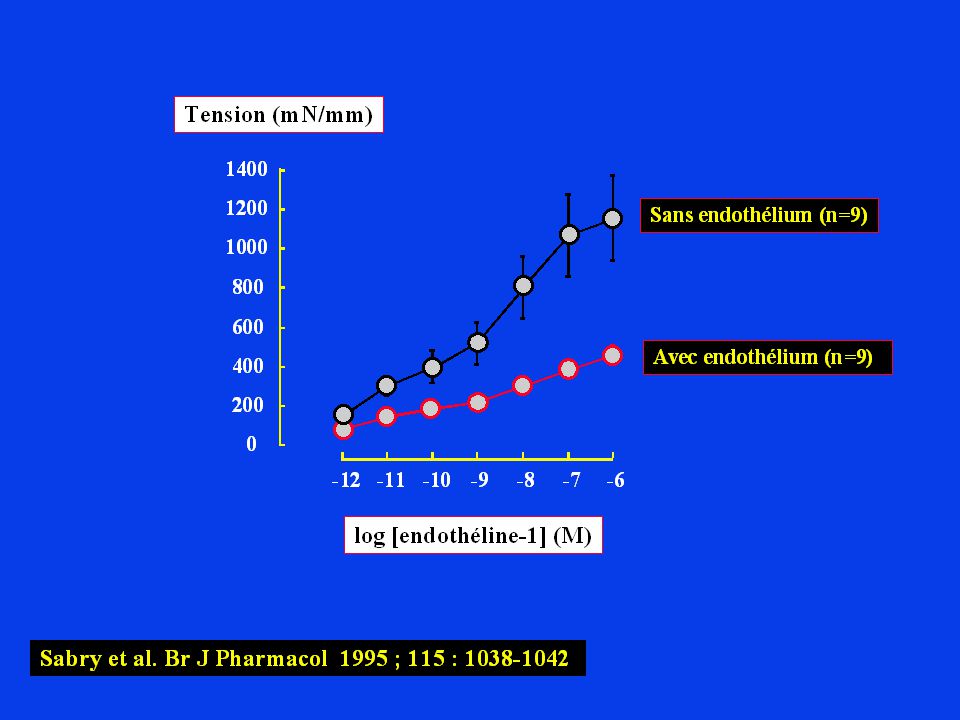
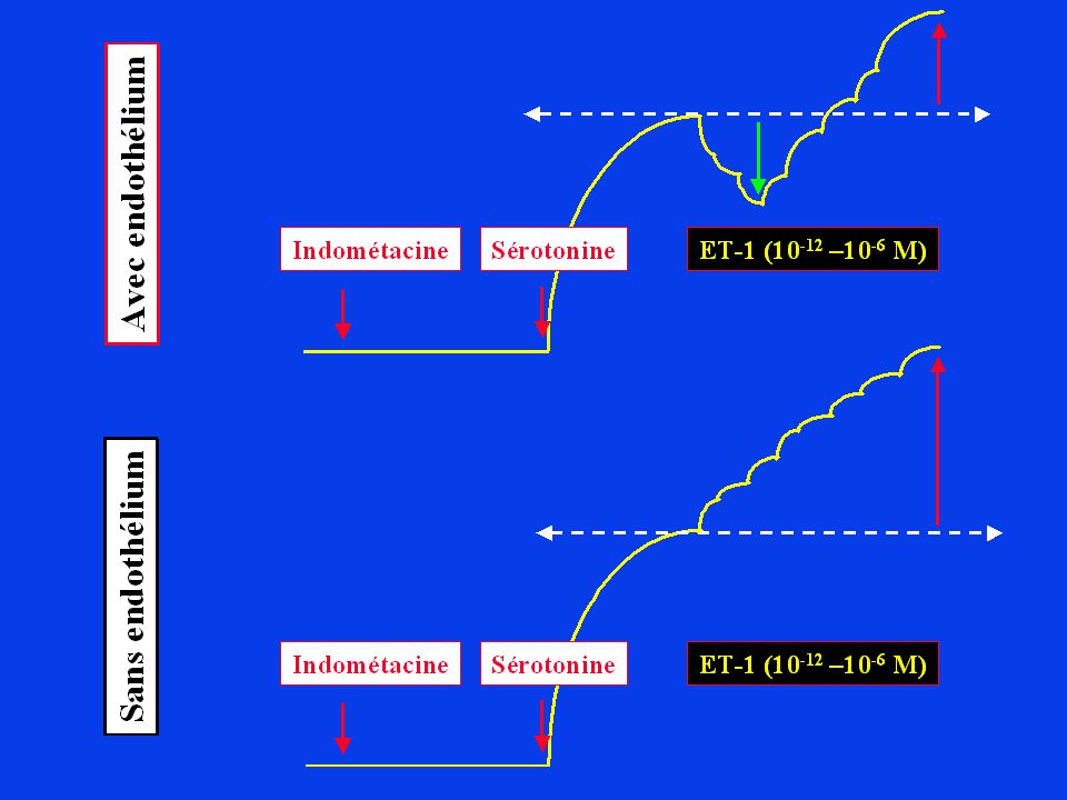
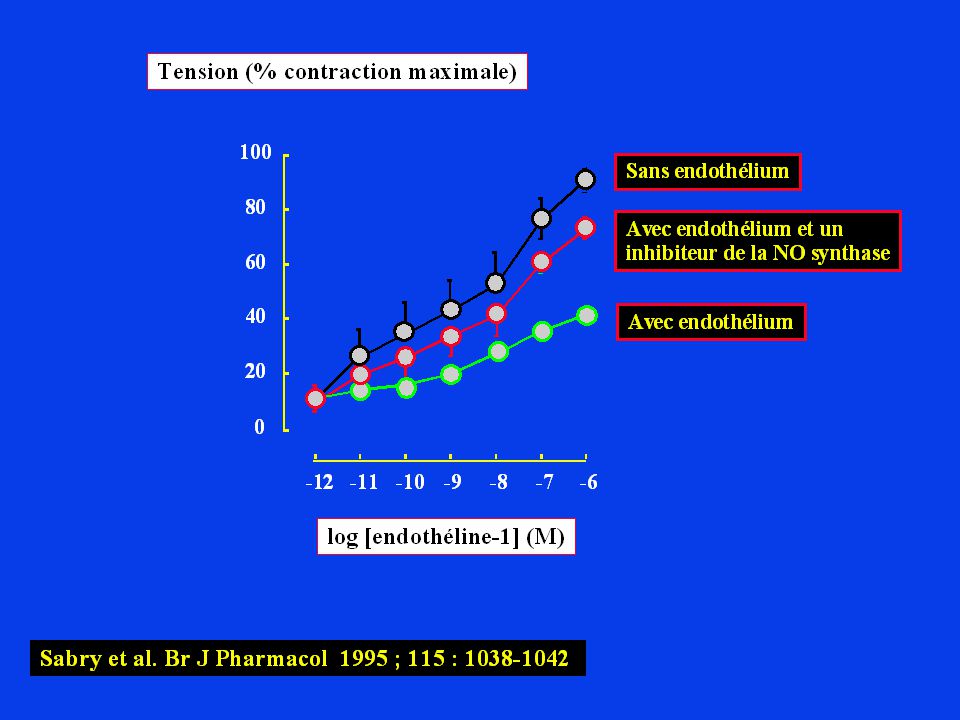
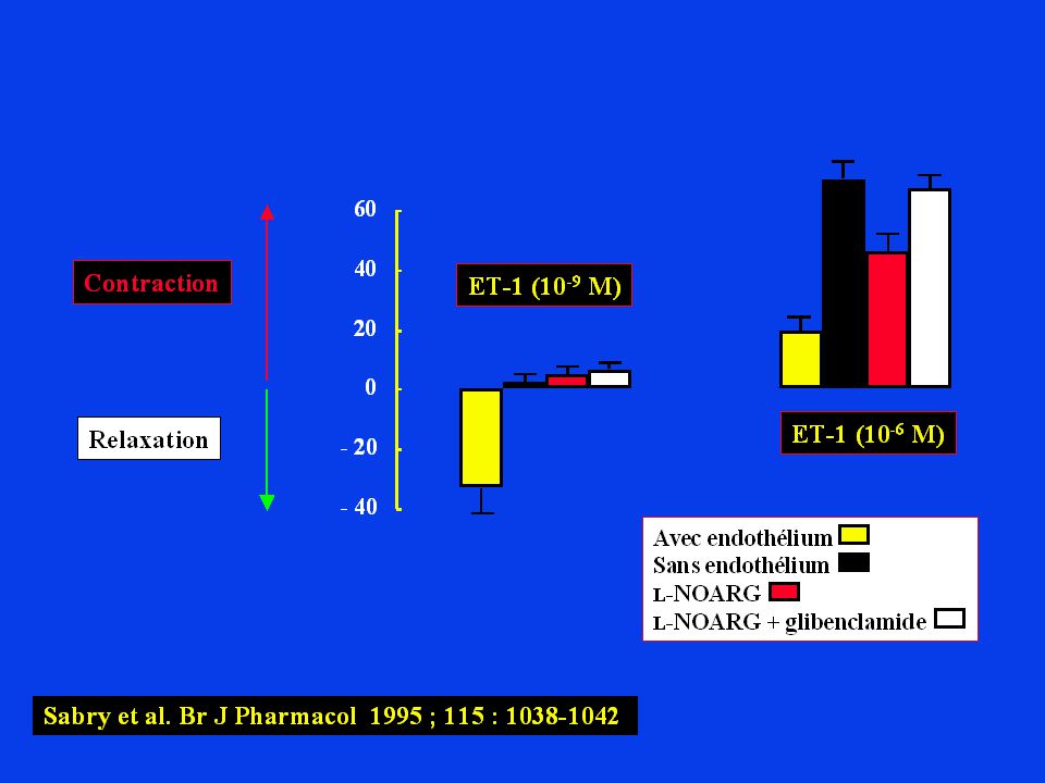
Endothelium-derived relaxing factor (EDRF)
Aorte de lapin sans endothélium Acétylcholine Phényléphrine Acétylcholine avec endothélium Phényléphrine Indométacine

8
Historique Furchgott RF, Zawadzki JV. The obligatory role of endothelial cells in the relaxation of arterial smooth muscle by acetylcholine. Nature 1980 ; 288 :

9
Endothélium Facteur endothélial Muscle lisse Relaxation

10
Guanylyl cyclase soluble
Shear stress 5-HT, NA, etc... ACh ADP Endothélium Ca2+ Synthase EDRF – Hémoglobine Dérivés oxygénés Muscle lisse GTP GMPc Guanylyl cyclase soluble Dérivés nitrés Relaxation

11
EDRF Relaxation Contraction Endothélium EDRF Muscle lisse ACh ACh Ca2+

12
Historique Furchgott RF, Zawadzki JV. The obligatory role of endothelial cells in the relaxation of arterial smooth muscle by acetylcholine. Nature 1980; 288: 373-6 Palmer RMJ, Ferrige AG, Moncada S. Nitric oxide release accounts for the biological activity of endothelium-derived relaxing factor. Nature 1987; 127:

13
Nitroglycerine, a 100 year old explosive and heart medicine
A New Principle Nitric Oxide, NO, is a short-lived, endogenously produced gas that acts as a signalling molecule in the body. Signal transmission by a gas, produced by one cell, which penetrates membranes and regulates the function of other cells is an entirely new principle for signalling in the human organism. Nitroglycerine, a 100 year old explosive and heart medicine In atherosclerosis, plaques reduce blood flow in the arteries. This decreases oxygen supply to the heart muscle causing chest pain (angina pectoris) and sometimes even myocardial infarction. Treatment with nitroglycerine provides NO, dilates the vessels, and increases blood flow. Thanks to this year's Nobel Laureates we now understand how nitroglycerine, an important heart medicine, works. It acts as a NO donor, causes dilation of the blood vessels, increases oxygen supply and protects the heart from damage and cell death.
 and sometimes even myocardial infarction. Treatment with nitroglycerine provides NO, dilates the vessels, and increases blood flow. Thanks to this year s Nobel Laureates we now understand how nitroglycerine, an important heart medicine, works. It acts as a NO donor, causes dilation of the blood vessels, increases oxygen supply and protects the heart from damage and cell death.")
14
Calmoduline Molecular Biology of the Cell, 3rd edn. Part III. Internal Organization of the Cell Chapter 15. Cell Signaling Signaling via G-Protein-linked Cell-Surface Receptors 11 Figure The structure of Ca2+/calmodulin based on x-ray diffraction and NMR studies. (A) The molecule has a "dumbbell" shape, with two globular ends connected by a long, exposed α helix. Each end has two Ca2+-binding domains, each with a loop of 12 amino acid residues in which aspartic acid and glutamic acid side chains form ionic bonds with Ca2+. The two Ca2+-binding sites in the carboxyl-terminal part of the molecule have a tenfold higher affinity for Ca2+ than those in the amino-terminal part. In solution the molecule is flexible, displaying a range of forms, from extended (as shown) to more compact. (B) The structural changes in Ca2+/calmodulin that occurs when it binds to a target protein (in this example a peptide that consists of the Ca2+/calmodulin-binding domain of a Ca2+/calmodulin-dependent protein kinase [myosin light-chain kinase, discussed below]). Note that the Ca2+/calmodulin has "jack-knifed" to surround the peptide. (A, based on x-ray crystallographic data from Y.S. Babu et al., Nature 315:37-40, © 1985 Macmillan Magazines Ltd.; B, based on x-ray crystallographic data from W.E. Meador, A.R. Means, and F.A. Quiocho, Science 257: , 1992, and on NMR data from M. Ikura et al., Science 256: , © 1992 the AAAS.) Molecular Biology of the Cell, 3rd edn. Part III. Internal Organization of the Cell Chapter 15. Cell Signaling Signaling via G-Protein-linked Cell-Surface Receptors
 The molecule has a dumbbell shape, with two globular ends connected by a long, exposed α helix. Each end has two Ca2+-binding domains, each with a loop of 12 amino acid residues in which aspartic acid and glutamic acid side chains form ionic bonds with Ca2+. The two Ca2+-binding sites in the carboxyl-terminal part of the molecule have a tenfold higher affinity for Ca2+ than those in the amino-terminal part. In solution the molecule is flexible, displaying a range of forms, from extended (as shown) to more compact. (B) The structural changes in Ca2+/calmodulin that occurs when it binds to a target protein (in this example a peptide that consists of the Ca2+/calmodulin-binding domain of a Ca2+/calmodulin-dependent protein kinase [myosin light-chain kinase, discussed below]). Note that the Ca2+/calmodulin has jack-knifed to surround the peptide. (A, based on x-ray crystallographic data from Y.S. Babu et al., Nature 315:37-40, © 1985 Macmillan Magazines Ltd.; B, based on x-ray crystallographic data from W.E. Meador, A.R. Means, and F.A. Quiocho, Science 257: , 1992, and on NMR data from M. Ikura et al., Science 256: , © 1992 the AAAS.) Molecular Biology of the Cell, 3rd edn. Part III. Internal Organization of the Cell Chapter 15. Cell Signaling Signaling via G-Protein-linked Cell-Surface Receptors.")
16
Vessels vivified by Akt acting on NO synthase Solomon H
Vessels vivified by Akt acting on NO synthase Solomon H. Snyder & Samie R. Jaffrey nature cell biology august 1999 volume 1 issue 4 pp E95-E96 Figure 1: Production of nitric oxide (NO) is usually thought to be triggered by the increases in cytosolic calcium (Ca2+) that occur following the stimulation of receptors for molecules such as acetylcholine and bradykinin. Ca2+ forms a complex with calmodulin, and together they bind to and activate endothelial nitric oxide synthase (eNOS), which produces NO. New results1, 2 indicate that important physiological triggers of NO production may actually activate eNOS through a Ca2+-independent pathway involving phosphatidylinositol-3-OH kinase (PI(3)K) and the serine/threonine kinase Akt. Activation of the PI(3)K/Akt pathway leads to eNOS phosphorylation and constitutive NO production. cGMP, cyclic GMP. Snyder & Jaffrey. Vessels vivified by Akt acting on NO synthase. Nature Cell Biology 1999; 1 : E95-E96.
 is usually thought to be triggered by the increases in cytosolic calcium (Ca2+) that occur following the stimulation of receptors for molecules such as acetylcholine and bradykinin. Ca2+ forms a complex with calmodulin, and together they bind to and activate endothelial nitric oxide synthase (eNOS), which produces NO. New results1, 2 indicate that important physiological triggers of NO production may actually activate eNOS through a Ca2+-independent pathway involving phosphatidylinositol-3-OH kinase (PI(3)K) and the serine/threonine kinase Akt. Activation of the PI(3)K/Akt pathway leads to eNOS phosphorylation and constitutive NO production. cGMP, cyclic GMP. Snyder & Jaffrey. Vessels vivified by Akt acting on NO synthase. Nature Cell Biology 1999; 1 : E95-E96.")
17
Am J Physiol Renal Physiol 280: F193-F206, 2001.
Fig. 3. The eNOS activation-deactivation cycle (1-8). eNOS, which is resident in the Golgi complex (anchored in the membrane by 1 myristoyl and 2 palmitoyl groups), is transported together with caveolin-1 to the caveolae at the plasma membrane (PM) via vesicles (1 and 2). Within the caveolae, eNOS is bound to caveolin-1, which inhibits eNOS activity. Shear stress signals via a potassium channel and the cytoskeleton, which results in tyrosine phosphorylation of specific proteins, activation of phosphatidylinositol 3-kinase, and subsequently in activation of Akt kinase. Akt activation by shear stress but also by VEGF activates eNOS by serine phosphorylation (PS; 3), which increases the affinity of eNOS for calmodulin. After agonist binding at the plasma membrane, eNOS-activating receptors translocate to caveolae. The 7-membrane-spanning domain-containing receptors activate G proteins, whereas the VEGF receptor signals via its tyrosine kinase domain. These receptors activate calcium channels at the caveolae. Furthermore, they activate calcium channels of the endoplasmic reticulum (ER) via phospholipase C and inositol 1,4,5-trisphosphate. This calcium flux induces binding of calmodulin to eNOS, whereas the eNOS-caveolin-1 interaction is disrupted. At the same time, eNOS is depalmitoylated (only the myristoyl moiety remains) and therefore translocates into the cytosol (4). On binding of calmodulin, eNOS generates NO (and under certain conditions superoxide, O2 ·), which may be enhanced by the interaction with Hsp90 (5). eNOS is inactivated by PS and by the dissociation of calmodulin (6). Perhaps phosphorylation induces the eNOS-calmodulin dissociation. After translocation to the Golgi complex (7), eNOS is repalmitoylated (8), which enables eNOS to be transported to the caveolae again. CAT-1, cationic amino acid transporter. Am J Physiol Renal Physiol 280: F193-F206, 2001.
. eNOS, which is resident in the Golgi complex (anchored in the membrane by 1 myristoyl and 2 palmitoyl groups), is transported together with caveolin-1 to the caveolae at the plasma membrane (PM) via vesicles (1 and 2). Within the caveolae, eNOS is bound to caveolin-1, which inhibits eNOS activity. Shear stress signals via a potassium channel and the cytoskeleton, which results in tyrosine phosphorylation of specific proteins, activation of phosphatidylinositol 3-kinase, and subsequently in activation of Akt kinase. Akt activation by shear stress but also by VEGF activates eNOS by serine phosphorylation (PS; 3), which increases the affinity of eNOS for calmodulin. After agonist binding at the plasma membrane, eNOS-activating receptors translocate to caveolae. The 7-membrane-spanning domain-containing receptors activate G proteins, whereas the VEGF receptor signals via its tyrosine kinase domain. These receptors activate calcium channels at the caveolae. Furthermore, they activate calcium channels of the endoplasmic reticulum (ER) via phospholipase C and inositol 1,4,5-trisphosphate. This calcium flux induces binding of calmodulin to eNOS, whereas the eNOS-caveolin-1 interaction is disrupted. At the same time, eNOS is depalmitoylated (only the myristoyl moiety remains) and therefore translocates into the cytosol (4). On binding of calmodulin, eNOS generates NO (and under certain conditions superoxide, O2 ·), which may be enhanced by the interaction with Hsp90 (5). eNOS is inactivated by PS and by the dissociation of calmodulin (6). Perhaps phosphorylation induces the eNOS-calmodulin dissociation. After translocation to the Golgi complex (7), eNOS is repalmitoylated (8), which enables eNOS to be transported to the caveolae again. CAT-1, cationic amino acid transporter. Am J Physiol Renal Physiol 280: F193-F206,")
18
Am J Physiol Renal Physiol 280: F193-F206, 2001.
Fig. 3. The eNOS activation-deactivation cycle (1-8). eNOS, which is resident in the Golgi complex (anchored in the membrane by 1 myristoyl and 2 palmitoyl groups), is transported together with caveolin-1 to the caveolae at the plasma membrane (PM) via vesicles (1 and 2). Within the caveolae, eNOS is bound to caveolin-1, which inhibits eNOS activity. Shear stress signals via a potassium channel and the cytoskeleton, which results in tyrosine phosphorylation of specific proteins, activation of phosphatidylinositol 3-kinase, and subsequently in activation of Akt kinase. Akt activation by shear stress but also by VEGF activates eNOS by serine phosphorylation (PS; 3), which increases the affinity of eNOS for calmodulin. After agonist binding at the plasma membrane, eNOS-activating receptors translocate to caveolae. The 7-membrane-spanning domain-containing receptors activate G proteins, whereas the VEGF receptor signals via its tyrosine kinase domain. These receptors activate calcium channels at the caveolae. Furthermore, they activate calcium channels of the endoplasmic reticulum (ER) via phospholipase C and inositol 1,4,5-trisphosphate. This calcium flux induces binding of calmodulin to eNOS, whereas the eNOS-caveolin-1 interaction is disrupted. At the same time, eNOS is depalmitoylated (only the myristoyl moiety remains) and therefore translocates into the cytosol (4). On binding of calmodulin, eNOS generates NO (and under certain conditions superoxide, O2 ·), which may be enhanced by the interaction with Hsp90 (5). eNOS is inactivated by PS and by the dissociation of calmodulin (6). Perhaps phosphorylation induces the eNOS-calmodulin dissociation. After translocation to the Golgi complex (7), eNOS is repalmitoylated (8), which enables eNOS to be transported to the caveolae again. CAT-1, cationic amino acid transporter. Am J Physiol Renal Physiol 280: F193-F206, 2001.
. eNOS, which is resident in the Golgi complex (anchored in the membrane by 1 myristoyl and 2 palmitoyl groups), is transported together with caveolin-1 to the caveolae at the plasma membrane (PM) via vesicles (1 and 2). Within the caveolae, eNOS is bound to caveolin-1, which inhibits eNOS activity. Shear stress signals via a potassium channel and the cytoskeleton, which results in tyrosine phosphorylation of specific proteins, activation of phosphatidylinositol 3-kinase, and subsequently in activation of Akt kinase. Akt activation by shear stress but also by VEGF activates eNOS by serine phosphorylation (PS; 3), which increases the affinity of eNOS for calmodulin. After agonist binding at the plasma membrane, eNOS-activating receptors translocate to caveolae. The 7-membrane-spanning domain-containing receptors activate G proteins, whereas the VEGF receptor signals via its tyrosine kinase domain. These receptors activate calcium channels at the caveolae. Furthermore, they activate calcium channels of the endoplasmic reticulum (ER) via phospholipase C and inositol 1,4,5-trisphosphate. This calcium flux induces binding of calmodulin to eNOS, whereas the eNOS-caveolin-1 interaction is disrupted. At the same time, eNOS is depalmitoylated (only the myristoyl moiety remains) and therefore translocates into the cytosol (4). On binding of calmodulin, eNOS generates NO (and under certain conditions superoxide, O2 ·), which may be enhanced by the interaction with Hsp90 (5). eNOS is inactivated by PS and by the dissociation of calmodulin (6). Perhaps phosphorylation induces the eNOS-calmodulin dissociation. After translocation to the Golgi complex (7), eNOS is repalmitoylated (8), which enables eNOS to be transported to the caveolae again. CAT-1, cationic amino acid transporter. Am J Physiol Renal Physiol 280: F193-F206,")
19
– + + CaMK II Am J Physiol Renal Physiol 280: F193-F206, 2001.
Fig. 2. Cellular events involved in the regulation of eNOS activity. The main pathway leading to a functional eNOS enzyme is depicted. For transcriptional regulation, (de)stabilization of eNOS mRNA, and protein-protein interactions, some examples of regulators of eNOS activity are shown. Once the enzyme is functional, the presence of substrate arginine and cofactor BH4 determines whether eNOS is producing nitric oxide (NO) or superoxide. EPO, erythropoietin; TNF-alpha , tumor necrosis factor-alpha ; LPS, lipopolysaccharide; VEGF, vascular endothelial growth factor; Hsp90, 90-kDa heat shock protein. + Am J Physiol Renal Physiol 280: F193-F206, 2001. CaMK II
stabilization of eNOS mRNA, and protein-protein interactions, some examples of regulators of eNOS activity are shown. Once the enzyme is functional, the presence of substrate arginine and cofactor BH4 determines whether eNOS is producing nitric oxide (NO) or superoxide. EPO, erythropoietin; TNF-alpha , tumor necrosis factor-alpha ; LPS, lipopolysaccharide; VEGF, vascular endothelial growth factor; Hsp90, 90-kDa heat shock protein. + Am J Physiol Renal Physiol 280: F193-F206, CaMK II.")
20
NO synthase inductible
NOSi ou NOS-2

21
NO synthases NOS constitutives inductible
Nature membranaire (E) soluble soluble (N) Quantité M M Durée secondes heures Corticoïdes insensible inhibition Calcium dépendant indépendant
 soluble. soluble (N) Quantité M 10-9M. Durée secondes heures. Corticoïdes insensible inhibition. Calcium dépendant indépendant.")
23
Transcription de la NOSi

26
Nathan & Shiloh. PNAS 2000; 97: 8841-8.
Nathan C, Shiloh MU. Reactive oxygen and nitrogen intermediates in the relationship between mammalian hosts and microbial pathogens. Proc. Natl. Acad. Sci. USA 2000; 97: , August 1. Fig. 1. ROI and RNI production in mammalian cells via phox and NOS: parallel but connecting paths. Nitroxyl anion (NO ), a one-electron reduction product of nitric oxide (·NO), is unlikely to arise from ·NO under physiologic conditions, but is considered by some investigators to be a primary and more toxic product of NOS (91). Reaction of RNI with cysteine sulfhydryls can lead either to S-nitrosylation or to oxidation to the sulfenic acid, as well as to disulfide bond formation (not shown), all of which are potentially reversible. Peroxynitrite anion (OONO ) and peroxynitrous acid (OONOH) have distinct patterns of reactivity (92), but for simplicity, the text refers to both as peroxynitrite. OONOH spontaneously decomposes via species resembling the reactive radicals, hydroxyl (OH·) and/or nitrogen dioxide (·NO2). When L-arginine is limiting, NOS can produce superoxide (O 2) along with ·NO, favoring the formation of peroxynitrite (5).
, a one-electron reduction product of nitric oxide (·NO), is unlikely to arise from ·NO under physiologic conditions, but is considered by some investigators to be a primary and more toxic product of NOS (91). Reaction of RNI with cysteine sulfhydryls can lead either to S-nitrosylation or to oxidation to the sulfenic acid, as well as to disulfide bond formation (not shown), all of which are potentially reversible. Peroxynitrite anion (OONO ) and peroxynitrous acid (OONOH) have distinct patterns of reactivity (92), but for simplicity, the text refers to both as peroxynitrite. OONOH spontaneously decomposes via species resembling the reactive radicals, hydroxyl (OH·) and/or nitrogen dioxide (·NO2). When L-arginine is limiting, NOS can produce superoxide (O 2) along with ·NO, favoring the formation of peroxynitrite (5).")
27
Molécules réagissant avec le NO
Métalloprotéines Hémoglobine Guanylyl cyclase soluble (effet dilatateur) Enzymes mitochondriales (effet cytotoxique) Dérivés oxygénés (O2–)
 Enzymes mitochondriales (effet cytotoxique) Dérivés oxygénés (O2–)")
28
NO synthase inductible
NOSi ou NOS-2

29
Macrophages Rappel chimique :
les nitrites (NO2–) et les nitrates (NO3–) sont des produits d’oxydation du NO. Historique Green et coll. (1981) : nitrates excrétés > nitrates ingérés Stuehr & Marletta (1985) : souris déficientes en macrophages excrètent peu de nitrates Hibbs et coll. (1987) : La L-arginine est la source de nitrates produits par les macrophages
 et les nitrates (NO3–) sont des produits d’oxydation du NO. Historique. Green et coll. (1981) : nitrates excrétés > nitrates ingérés. Stuehr & Marletta (1985) : souris déficientes en macrophages excrètent peu de nitrates. Hibbs et coll. (1987) : La L-arginine est la source de nitrates produits par les macrophages.")
30
Macrophages Les macrophages activés sont capables de synthétiser du NO à partir de la L-arginine. Remarque : les nitrates mesurés ne sont que des produits d’oxydation du NO. L-arginine NOSi NO. NO2- NO3- nitrites nitrates

32
Pourquoi Mr Hyde ? Quantité de NO (fonction de la NOS)
Qualité de l’environnement (réactivité du NO)
")
33
Nathan & Shiloh. PNAS 2000; 97: 8841-8.
Nathan C, Shiloh MU. Reactive oxygen and nitrogen intermediates in the relationship between mammalian hosts and microbial pathogens. Proc. Natl. Acad. Sci. USA 2000; 97: , August 1. Fig. 1. ROI and RNI production in mammalian cells via phox and NOS: parallel but connecting paths. Nitroxyl anion (NO ), a one-electron reduction product of nitric oxide (·NO), is unlikely to arise from ·NO under physiologic conditions, but is considered by some investigators to be a primary and more toxic product of NOS (91). Reaction of RNI with cysteine sulfhydryls can lead either to S-nitrosylation or to oxidation to the sulfenic acid, as well as to disulfide bond formation (not shown), all of which are potentially reversible. Peroxynitrite anion (OONO ) and peroxynitrous acid (OONOH) have distinct patterns of reactivity (92), but for simplicity, the text refers to both as peroxynitrite. OONOH spontaneously decomposes via species resembling the reactive radicals, hydroxyl (OH·) and/or nitrogen dioxide (·NO2). When L-arginine is limiting, NOS can produce superoxide (O 2) along with ·NO, favoring the formation of peroxynitrite (5).
, a one-electron reduction product of nitric oxide (·NO), is unlikely to arise from ·NO under physiologic conditions, but is considered by some investigators to be a primary and more toxic product of NOS (91). Reaction of RNI with cysteine sulfhydryls can lead either to S-nitrosylation or to oxidation to the sulfenic acid, as well as to disulfide bond formation (not shown), all of which are potentially reversible. Peroxynitrite anion (OONO ) and peroxynitrous acid (OONOH) have distinct patterns of reactivity (92), but for simplicity, the text refers to both as peroxynitrite. OONOH spontaneously decomposes via species resembling the reactive radicals, hydroxyl (OH·) and/or nitrogen dioxide (·NO2). When L-arginine is limiting, NOS can produce superoxide (O 2) along with ·NO, favoring the formation of peroxynitrite (5).")
34
Transcription de la NOSi

35
Figure 4. Some anti-inflammatory effects are likely to be mediated by inhibition of NF- B, which is activated by many stimuli that lead to exacerbations of asthma and leads to the expression of multiple genes that are abnormally expressed in asthmatic airways. Barnes et al. Am J Respir Crit Care Med 1998; 157: S1-S53.

36
NO Stimulus inflammatoire Réponse inflammatoire Cytokines Noyau I-kB
GC REC GC Noyau p50 p65 NF-kB Gène I-kB I-kB GRE NOSi ARNm Élément de réponse kB p50 p65 TATA Gène NOSi

37
NO synthase neuronale NOSn ou NOS-1

38
Garthwaite et al. Nature 1988; 336: 385-8.
N-méthyl-D-Aspartate Aorte de rat sans endothélium Phényléphrine Phényléphrine + neurones du cervelet NMDA NMDA + Hb

40
Voie nerveuse nitroxidergique
Innervation des muscles lisses (ML) ML vasculaire : vasorelaxation ML bronchique : bronchodilatation ML digestif : relâchement musculaire et sphinctérien Corps caverneux : fonction érectile
 ML vasculaire : vasorelaxation. ML bronchique : bronchodilatation. ML digestif : relâchement musculaire et sphinctérien Corps caverneux : fonction érectile.")
41
NO et physiologie vasculaire
Endothélium vasculaire NO Muscle lisse vasculaire - + GMPc Relaxation Prolifération Innervation autonome (NANCi) NO
 NO.")
42
(Ca2+)i NO t24 AMPA calmoduline NMDA CaM kinase II L-Glu NO Synthase
GMPc L-Glu calmoduline Ca2+ Ca2+ NMDA NO Synthase NO t24

43
NO synthases (NOS) NOS-1 (neuronal) 150 kDa 12q24.2 FMN FAD NADPH Hème
NH2 COOH Calmoduline NOS-2 (macrophagique) 130 kDa 17cen-q12 FMN FAD NADPH Hème NH2 COOH acide myristique NOS-3 (endothéliale) 135 kDa 7q35-36 FMN FAD NADPH P Hème NH2 COOH
 130 kDa. 17cen-q12. FMN. FAD. NADPH. Hème. NH2. COOH. acide. myristique. NOS-3 (endothéliale) 135 kDa. 7q FMN. FAD. NADPH. P. Hème. NH2. COOH.")
44
NO synthase (NOS) Nature Reviews Drug Discovery 1; (2002); doi: /nrd960 BLOCKING NO SYNTHESIS: HOW, WHERE AND WHY? Figure 1 | The NOS pathway. For enzymatic activity, nitric oxide synthase (NOS) enzymes must dimerize and bind the cofactors tetrahydrobiopterin (BH4), haem, flavin mononucleotide (FMN) and flavin adenine dinucleotide (FAD). On binding calmodulin (CAL), the active enzyme catalyses the oxidation of L-arginine to citrulline and nitric oxide (NO) and requires molecular oxygen and NADPH as co-substrates. Each NOS dimer coordinates a single zinc (Zn) atom.
 enzymes must dimerize and bind the cofactors tetrahydrobiopterin (BH4), haem, flavin mononucleotide (FMN) and flavin adenine dinucleotide (FAD). On binding calmodulin (CAL), the active enzyme catalyses the oxidation of L-arginine to citrulline and nitric oxide (NO) and requires molecular oxygen and NADPH as co-substrates. Each NOS dimer coordinates a single zinc (Zn) atom.")
45
– – Muscle lisse Endothélium Relaxation GCs Prolifération NO NO O2 ACh
ADP L-arginine Ca2+ Endothélium G O2 Ca2+ L-NAME L-NMMA Calmoduline NOS – NADPH L-arginine L-citrulline NO GTP NO Relaxation GCs Ca2+ GMPc Muscle lisse Prolifération –

46
Muscle lisse vasculaire Relaxation
NO Inhibiteurs de la PDE-5 Muscle lisse vasculaire GTP GMPc GCs PDE-5 5’-GMP PKG Relaxation

47
Les guanylates cyclases
Lucas et al. Pharmacol Rev 2000; 52: Peptides natriurétiques (ANP, BNP) Guanyline Toxines (STa)
 Guanyline. Toxines (STa)")
48
Lucas et al. Pharmacol Rev 2002
Sindić &Schlatter. Pflügers Arch 2005

49
Ca2+ 10-3 M 10-7 à 10-5 M IP3 gradient 10-2 M réticulum endoplasmique
échangeur Na+/Ca2+ gradient canaux calciques Ca2+ ATPase IP3 récepteur-canal Ca2+ ATPase réticulum endoplasmique

50
Kimberly A. Lucas, Giovanni M
Kimberly A. Lucas, Giovanni M. Pitari, Shiva Kazerounian, Inez Ruiz-Stewart, Jason Park, Stephanie Schulz, Kenneth P. Chepenik and Scott A. Waldman Pharmacol Rev Vol. 52, Issue 3, , September 2000 Fig. 4. Molecular mechanisms underlying vascular smooth muscle relaxation mediated by cyclic GMP. Cyclic GMP induces smooth muscle relaxation by reducing [Ca2+]i and desensitizing the contractile apparatus to Ca2+. Cyclic GMP reduces [Ca2+]i by (1) inhibiting Ca2+ influx through L-type Ca2+ channels; (2) increasing Ca2+ efflux through activation of (2d) the Ca2+-pumping ATPase and (2b) the Na+/Ca2+ exchanger; also, cGMP may produce membrane hyperpolarization through activation of (2c) the Na+/K+ ATPase and (2a) K+ channels, thereby increasing Ca2+ extrusion by the Na+/Ca2+ exchanger; (3) increasing of Ca2+ sequestration through activation of the sarcoplasmic reticulum Ca2+-pumping ATPase [Ph, phospholamban]; and (4) decreasing of Ca2+ mobilization through inhibition of agonist-induced IP3 formation or inhibition of the IP3 receptor in the sarcoplasmic reticulum. R, receptor; G, G protein; PLC, phospholipase C; IP3R, IP3 receptor. Cyclic GMP desensitizes the contractile apparatus to Ca2+ (5) probably by activating myosin light chain phosphatase, resulting in dephosphorylation of the 20 kDa myosin light chain. Lucas et al. Pharmacol Rev 2000; 52:
 inhibiting Ca2+ influx through L-type Ca2+ channels; (2) increasing Ca2+ efflux through activation of (2d) the Ca2+-pumping ATPase and (2b) the Na+/Ca2+ exchanger; also, cGMP may produce membrane hyperpolarization through activation of (2c) the Na+/K+ ATPase and (2a) K+ channels, thereby increasing Ca2+ extrusion by the Na+/Ca2+ exchanger; (3) increasing of Ca2+ sequestration through activation of the sarcoplasmic reticulum Ca2+-pumping ATPase [Ph, phospholamban]; and (4) decreasing of Ca2+ mobilization through inhibition of agonist-induced IP3 formation or inhibition of the IP3 receptor in the sarcoplasmic reticulum. R, receptor; G, G protein; PLC, phospholipase C; IP3R, IP3 receptor. Cyclic GMP desensitizes the contractile apparatus to Ca2+ (5) probably by activating myosin light chain phosphatase, resulting in dephosphorylation of the 20 kDa myosin light chain. Lucas et al. Pharmacol Rev 2000; 52:")
51
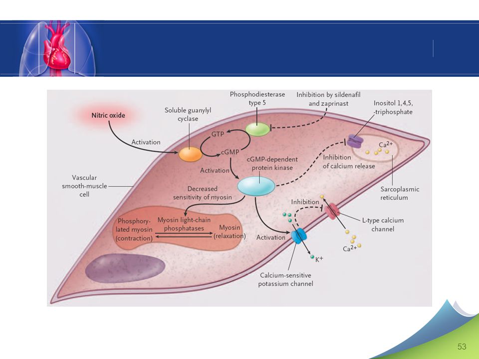
Canaux K+ et Ca2+ Am. J. Respir. Crit. Care Med., Volume 157, Number 4, April 1998, S101-S108 The Pulmonary Circulation Snapshots of Progress JOHN T. REEVES and LEWIS J. RUBIN Figure 1. The effect of redox state on membrane potassium channel activity. Normoxia is associated with open channels, whereas hypoxia results in closing of the potassium channels, leading to membrane depolarization, opening of voltage-gated calcium channels, influx of calcium into the cytosol, and vasoconstriction. Oxidizing compounds mimic normoxia, while reducing agents mimic the effects of hypoxia. Reeves & Rubin. Am J Respir Crit Care Med 1998; 157: S101-8.

52
GMPc et canaux potassiques
Potassium Channel Function in Vascular Disease Christopher G. Sobey Arteriosclerosis, Thrombosis, and Vascular Biology. 2001;21: Figure 2. Some potential mechanisms involving K+ channel–mediated, endothelium-dependent hyperpolarization. Stimulation of endothelial cell receptors by agonists such as acetylcholine (ACh), bradykinin (BK), and substance P (SP) may cause release of several endothelium-derived relaxing factors. These include NO, EDHF(s), and prostacyclin (PGI2), each of which can induce relaxation of underlying vascular muscle through activation of K+ channels. In the case of NO and PGI2, this may involve the intracellular accumulation of a second messenger (cyclic GMP and cAMP, respectively). Like EDHF, NO may activate K+ channels directly. The production of EDHF may depend on the bioavailability of NO, such that EDHF release may be more significant under conditions in which NO production is impaired. The hyperpolarization occurring in response to K+ channel activation leads to vasodilatation, as described in Figure 1 . Because all 3 endothelial factors may normally activate vascular K+ channels, endothelial dysfunction occurring during cardiovascular disease states may result in vascular depolarization and/or abnormal K+ channel function, leading to increased vascular tone and reduced blood flow. Sobey. Arterio Scler Thromb Vasc Biol 2001; 21:
, bradykinin (BK), and substance P (SP) may cause release of several endothelium-derived relaxing factors. These include NO, EDHF(s), and prostacyclin (PGI2), each of which can induce relaxation of underlying vascular muscle through activation of K+ channels. In the case of NO and PGI2, this may involve the intracellular accumulation of a second messenger (cyclic GMP and cAMP, respectively). Like EDHF, NO may activate K+ channels directly. The production of EDHF may depend on the bioavailability of NO, such that EDHF release may be more significant under conditions in which NO production is impaired. The hyperpolarization occurring in response to K+ channel activation leads to vasodilatation, as described in Figure 1 . Because all 3 endothelial factors may normally activate vascular K+ channels, endothelial dysfunction occurring during cardiovascular disease states may result in vascular depolarization and/or abnormal K+ channel function, leading to increased vascular tone and reduced blood flow. Sobey. Arterio Scler Thromb Vasc Biol 2001; 21:")
54
Friebe & Koesling. Circ Res 2003; 93: 96-105.

55
Les isoformes des phosphodiestérases
Cyclic GMP Phosphodiesterases and Regulation of Smooth Muscle Function Sergei D. Rybalkin, Chen Yan, Karin E. Bornfeldt, Joseph A. Beavo Circulation Research. 2003;93:280 Figure 1. Domain organization of 11 PDE gene families. All PDEs share significant homology in their catalytic domain but differ greatly in their N-terminal parts, which contain different types of regulatory domains. UCR domain is an upstream conserved region found only in PDE4. GAF and PAS domains are derived from the first letters of initial members of their corresponding groups. GAF domains (cGMP-regulated phosphodiesterases, several adenylyl cyclases, and FhlA) were originally defined in PDE2, PDE5, and PDE6 and later shown in PDE10 and PDE11. PAS domain (Per, ARNT, and Sim proteins) was found only in PDE8. Rybalkin et al. Circ Res 2003; 93: 280.
 were originally defined in PDE2, PDE5, and PDE6 and later shown in PDE10 and PDE11. PAS domain (Per, ARNT, and Sim proteins) was found only in PDE8. Rybalkin et al. Circ Res 2003; 93: 280.")
56
Figure 5. PDE5 is directly activated upon cGMP binding to its GAF A domain. Without cGMP bound, PDE5 is in a nonactivated state. PDE5 is converted into an activated state after binding of cGMP to the GAF A domain. Only activated PDE5 is phosphorylated by PKG. Adapted from Rybalkin SD, Rybalkina IG, Shimizu-Albergine M, Tang X-B, Beavo JA. PDE5 is converted to an activated state upon cGMP binding to the GAF A domain. EMBO J. 2003;22:469–478. From: Rybalkin: Circ Res, Volume 93(4).August 22, Rybalkin et al. Circ Res 2003; 93: 280.

57
GMPc : un jeune second (messager) de 43 ans
1963 : dosage urinaire du GMPc (Ashman et al.) 1969 : identification de la guanylate cyclase (Hardman & Sutherland) 1980 : découverte de l’EDRF (Furchgott & Zawadzki) 1984 : activation de la guanylate cyclase membranaire par l’ANP (Waldman et al.) 1987 : identification du NO (EDRF) (Palmer et al., Ignarro et al.) 1998 : Prix Nobel accordé à Furchgott, Ignarro et Murad.
 1969 : identification de la guanylate cyclase (Hardman & Sutherland) 1980 : découverte de l’EDRF (Furchgott & Zawadzki) 1984 : activation de la guanylate cyclase membranaire par l’ANP (Waldman et al.) 1987 : identification du NO (EDRF) (Palmer et al., Ignarro et al.) 1998 : Prix Nobel accordé à Furchgott, Ignarro et Murad.")
58
GCs Relaxation Endothélium Muscle lisse PDE5 vasculaire PKG ACh ADP
NOS O2 Ca2+ L-arginine ACh ADP G NADPH Calmoduline L-citrulline NO GTP GMPc Relaxation Muscle lisse vasculaire GCs PKG Ca2+ 5’-GMP PDE5 Donneurs de NO Inhibiteurs de la PDE5 YC1 ODQ KT-5823

59
Molécules réagissant avec le NO
Métalloprotéines Hémoglobine Guanylate cyclase soluble (effet stimulant, dilatateur) Enzymes mitochondriales (effet inhibiteur, cytotoxique) Dérivés oxygénés (O2–)
 Enzymes mitochondriales (effet inhibiteur, cytotoxique) Dérivés oxygénés (O2–)")
60
NO ONOO– a b GMPc O2– Caspases NF-kB PDE ras Canaux ioniques PKG GTP
Anion peroxynitrite O2– Anion superoxyde NO Guanylate Cyclase Soluble a b GTP GMPc NF-kB ras GDP GTP Caspases PKG Canaux ioniques PDE AMPc GMP

61
Facteurs de transcription
NO. LIPIDES PROTEINES Enzymes Facteurs de transcription (NF-kB, AP-1) Hème Fe Hb, GCs, catalase, cytochromes Fe-S aconitase -SH S-nitrosylation Tyrosine Nitration des résidus tyrosyls
 Hème. Fe. Hb, GCs, catalase, cytochromes. Fe-S. aconitase. -SH. S-nitrosylation. Tyrosine. Nitration des résidus tyrosyls.")
62
The pathfinders Beavo & Brunton. Nature Rev Mol Cell Biol 2002; 3:

63
(kieselghur + nitroglycérine)
Thomas Lauder Brunton (Lancet 1867; 2: 97-98) Alfred Nobel ( ) R.F. Furchgott 1980 EDRF Dynamite (kieselghur + nitroglycérine) Furchgott, Ignarro & Moncada 1987 NO = EDRF = dérivé nitré endogène Fondation NOBEL Prix NOBEL 1998
 Alfred Nobel. ( ) R.F. Furchgott EDRF. Dynamite. (kieselghur + nitroglycérine) Furchgott, Ignarro & Moncada NO = EDRF = dérivé nitré endogène. Fondation NOBEL. Prix NOBEL")
64
Endothélines Master M1 18 février 2006
Pr A.T. Dinh-Xuan Laboratoire de Physiologie - UPRES-EA 2511 Faculté de Médecine, Université Paris 5 René Desartes

65
Targets for Current or Emerging Therapies in Pulmonary Arterial Hypertension
Figure 1. Targets for Current or Emerging Therapies in Pulmonary Arterial Hypertension. Three major pathways involved in abnormal proliferation and contraction of the smooth-muscle cells of the pulmonary artery in patients with pulmonary arterial hypertension are shown. These pathways correspond to important therapeutic targets in this condition and play a role in determining which of four classes of drugs — endothelin-receptor antagonists, nitric oxide, phosphodiesterase type 5 inhibitors, and prostacyclin derivatives — will be used. At the top of the figure, a transverse section of a small pulmonary artery (<500 µm in diameter) from a patient with severe pulmonary arterial hypertension shows intimal proliferation and marked medial hypertrophy. Dysfunctional pulmonary-artery endothelial cells (blue) have decreased production of prostacyclin and endogenous nitric oxide, with an increased production of endothelin-1 — a condition promoting vasoconstriction and proliferation of smooth-muscle cells in the pulmonary arteries (red). Current or emerging therapies interfere with specific targets in smooth-muscle cells in the pulmonary arteries. In addition to their actions on smooth-muscle cells, prostacyclin derivatives and nitric oxide have several other properties, including antiplatelet effects. Plus signs denote an increase in the intracellular concentration; minus signs blockage of a receptor, inhibition of an enzyme, or a decrease in the intracellular concentration; and cGMP cyclic guanosine monophosphate. Humbert, M. et al. N Engl J Med 2004;351:
 from a patient with severe pulmonary arterial hypertension shows intimal proliferation and marked medial hypertrophy. Dysfunctional pulmonary-artery endothelial cells (blue) have decreased production of prostacyclin and endogenous nitric oxide, with an increased production of endothelin-1 — a condition promoting vasoconstriction and proliferation of smooth-muscle cells in the pulmonary arteries (red). Current or emerging therapies interfere with specific targets in smooth-muscle cells in the pulmonary arteries. In addition to their actions on smooth-muscle cells, prostacyclin derivatives and nitric oxide have several other properties, including antiplatelet effects. Plus signs denote an increase in the intracellular concentration; minus signs blockage of a receptor, inhibition of an enzyme, or a decrease in the intracellular concentration; and cGMP cyclic guanosine monophosphate. Humbert, M. et al. N Engl J Med 2004;351:")
66
2 4 5 6 7 14 6 7 Fig. 1. Amino acid sequence of human ETs and STXc. Amino acid substitutions with respect to ET-1 are indicated. Pharmacological Reviews. Vol. 51, Issue 3, , September 1999 Autocrine-Paracrine Endothelin System in the Physiology and Pathology of Steroid-Secreting Tissues Gastone G. Nussdorfer1, Gian Paolo Rossi, Ludwik K. Malendowicz and Giuseppina Mazzocchi

67
Fig. 1. Amino acid sequence of human ETs and STXc
Fig. 1. Amino acid sequence of human ETs and STXc. Amino acid substitutions with respect to ET-1 are indicated. Pharmacological Reviews. Vol. 51, Issue 3, , September 1999 Autocrine-Paracrine Endothelin System in the Physiology and Pathology of Steroid-Secreting Tissues Gastone G. Nussdorfer1, Gian Paolo Rossi, Ludwik K. Malendowicz and Giuseppina Mazzocchi

68
Fig. 2. Biosynthesis and processing of ET-1 from ppET-1
Fig. 2. Biosynthesis and processing of ET-1 from ppET-1. SP, signal peptide. Pharmacological Reviews. Vol. 51, Issue 3, , September 1999 Autocrine-Paracrine Endothelin System in the Physiology and Pathology of Steroid-Secreting Tissues Gastone G. Nussdorfer1, Gian Paolo Rossi, Ludwik K. Malendowicz and Giuseppina Mazzocchi

70
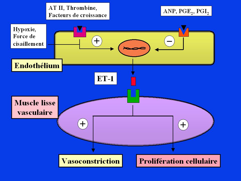
Fig. 1. A schema showing possible regulatory mechanisms of endothelin-1 production in various cells. AP-1, transcription factor; DG, 1,2-diacylglycerol; G, GTP-binding protein; GRB2, growth factor receptor bound 2; Ins(1,4,5)P3, inositol (1,4,5)-trisphosphate; MAPK, mitogen-activated protein kinase; MEK, MAPK/ERK (extracellular signal-regulated protein kinase) kinase; NF-1, transcription factor; PI3K, phosphatidylinositol-3 kinase; PIP2, phosphatidylinositol (4,5)-bisphosphate; PLC, phospholipase C; PKC, protein kinase C; PYK2, proline-rich tyrosine kinase 2; SHC, Sh2-containing protein; SOS, son of sevenless; SR, sarcoplasmic reticulum; TGF- , transforming growth factor ; TPA, 12-o-tetradecanoylphorbol 13-acetate. Heart failure and endothelin receptor antagonists Takashi Miyauchi a and Katsutoshi Goto b Trends in Pharmacological Sciences 1999, 20:
P3, inositol (1,4,5)-trisphosphate; MAPK, mitogen-activated protein kinase; MEK, MAPK/ERK (extracellular signal-regulated protein kinase) kinase; NF-1, transcription factor; PI3K, phosphatidylinositol-3 kinase; PIP2, phosphatidylinositol (4,5)-bisphosphate; PLC, phospholipase C; PKC, protein kinase C; PYK2, proline-rich tyrosine kinase 2; SHC, Sh2-containing protein; SOS, son of sevenless; SR, sarcoplasmic reticulum; TGF- , transforming growth factor ; TPA, 12-o-tetradecanoylphorbol 13-acetate. Heart failure and endothelin receptor antagonists Takashi Miyauchi a and Katsutoshi Goto b Trends in Pharmacological Sciences 1999, 20:")
77
Glycémie KATP K+ Cellule b Ca2+ Ca (VOC) I ATP I Dépolarisation Ca2+
Glucose Ouvreurs des KATP Sulfamides hypoglycémiants K+ KATP ATP Dépolarisation I I Ca2+ Ca2+ Ca (VOC)
")
79
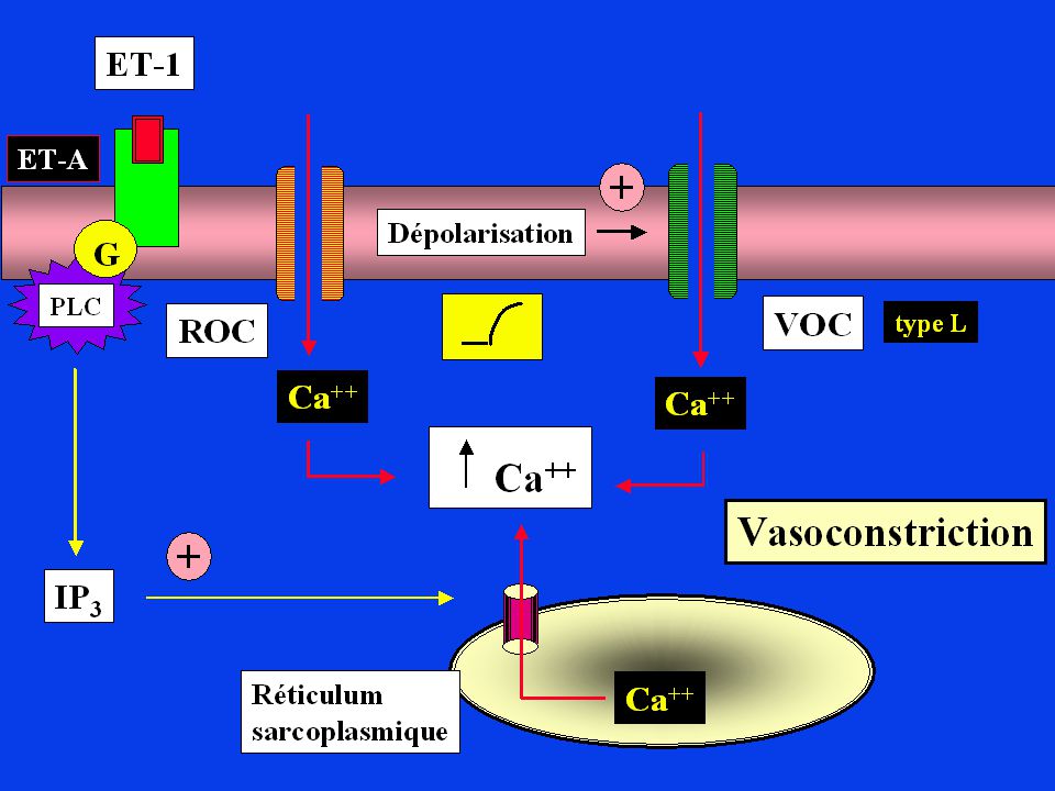
A B C Nature Reviews Drug Discovery 1; (2002); doi: /nrd962 NEW THERAPEUTICS THAT ANTAGONIZE ENDOTHELIN: PROMISES AND FRUSTRATIONS Figure 1 | Regulation of ET-1 synthesis, pathway of ET generation and ET-receptor-mediated actions on smooth muscle cells. Endothelin-1 (ET-1) synthesis is regulated by many factors; stimulators are highlighted in green, and inhibitors are highlighted in red. The product of ET1 transcription is prepro-ET-1, which is cleaved by a neutral endopeptidase to form the active precursor pro-ET-1 or big ET-1. Big ET-1 is converted to the mature peptide by the metalloproteinase endothelin-converting enzyme-1 (ECE-1)1. Two ET receptors have been identified in the vasculature: ET type-A receptors (ETA) reside in vascular smooth muscle cells and mediate vasoconstriction and cell proliferation, whereas ETB receptors reside on endothelial cells and are mainly vasodilatory through NO (which in turn can mediate the anti-apoptotic effects of ET-1), and regulate the synthesis of ET-1. ETB receptors on smooth muscle cells can elicit vessel contraction43. CsA, cyclosporin A; EGF, epidermal growth factor; HGF, hepatocyte growth factor; IL-1, interleukin-1; LDL, low-density lipoprotein; VEGF, vascular endothelial growth factor.
 synthesis is regulated by many factors; stimulators are highlighted in green, and inhibitors are highlighted in red. The product of ET1 transcription is prepro-ET-1, which is cleaved by a neutral endopeptidase to form the active precursor pro-ET-1 or big ET-1. Big ET-1 is converted to the mature peptide by the metalloproteinase endothelin-converting enzyme-1 (ECE-1)1. Two ET receptors have been identified in the vasculature: ET type-A receptors (ETA) reside in vascular smooth muscle cells and mediate vasoconstriction and cell proliferation, whereas ETB receptors reside on endothelial cells and are mainly vasodilatory through NO (which in turn can mediate the anti-apoptotic effects of ET-1), and regulate the synthesis of ET-1. ETB receptors on smooth muscle cells can elicit vessel contraction43. CsA, cyclosporin A; EGF, epidermal growth factor; HGF, hepatocyte growth factor; IL-1, interleukin-1; LDL, low-density lipoprotein; VEGF, vascular endothelial growth factor.")
80
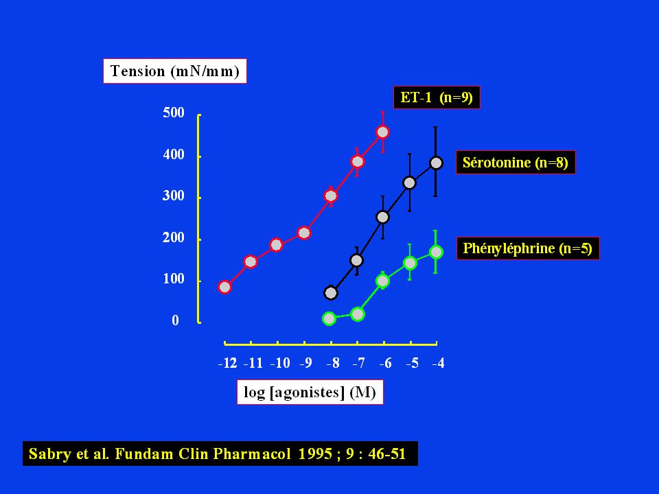
Lüscher & Barton. Circulation 2000; 102: 2434-40
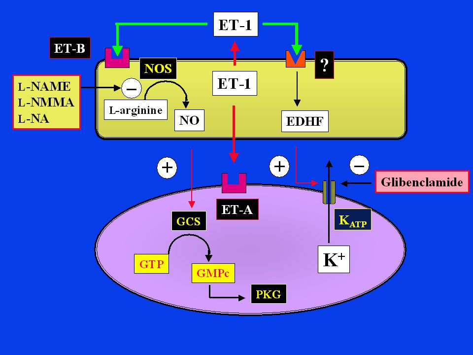
Figure 3. Vascular effects of ET-1. ET-1 is generated in endothelial and smooth muscle cells in response to oxidized LDL, angiotensin II (Ang II), etc. The stimulation of endothelial ETB receptors increases the release of NO, whereas ETA receptors mediate contraction and cell proliferation and migration. ET-1 stimulates interleukin (IL) and tumor necrosis factor- (TNF ) expression in monocytes, leukocyte adherence, platelet aggregation, and adhesion molecule expression. ET-1 stimulates the production and action of growth factors, DNA and protein synthesis, and cell cycle progression. ONOO- indicates peroxynitrite; NOS, nitric oxide synthase; MCP-1, monocyte chemoattractant protein-1; ICAM-1, intracellular adhesion molecule-1; VCAM-1, vascular cell adhesion molecule-1; oxLDL, oxidized low density lipoprotein; O2-, superoxide anion; LOX, lectin-like oxidized LDL receptor; TGF ß-1, transforming growth factor-ß1; NADPHox, nicotinamide adenine dinucleotide phosphate oxidase; PAI-1, plasminogen activator inhibitor-1; VEGF, vascular endothelial growth factor; bFGF-2, basic fibroblast growth factor-2; PDGF, platelet-derived growth factor; +, stimulation; and -, inhibition. Endothelins and Endothelin Receptor Antagonists Therapeutic Considerations for a Novel Class of Cardiovascular Drugs Presented in part at the 71st Scientific Sessions of the American Heart Association, Dallas, Tex, November 9–12, 1998. Thomas F. Lüscher, MD; Matthias Barton, MD (Circulation. 2000;102: ) © 2000 American Heart Association, Inc. Lüscher & Barton. Circulation 2000; 102:
, etc. The stimulation of endothelial ETB receptors increases the release of NO, whereas ETA receptors mediate contraction and cell proliferation and migration. ET-1 stimulates interleukin (IL) and tumor necrosis factor- (TNF ) expression in monocytes, leukocyte adherence, platelet aggregation, and adhesion molecule expression. ET-1 stimulates the production and action of growth factors, DNA and protein synthesis, and cell cycle progression. ONOO- indicates peroxynitrite; NOS, nitric oxide synthase; MCP-1, monocyte chemoattractant protein-1; ICAM-1, intracellular adhesion molecule-1; VCAM-1, vascular cell adhesion molecule-1; oxLDL, oxidized low density lipoprotein; O2-, superoxide anion; LOX, lectin-like oxidized LDL receptor; TGF ß-1, transforming growth factor-ß1; NADPHox, nicotinamide adenine dinucleotide phosphate oxidase; PAI-1, plasminogen activator inhibitor-1; VEGF, vascular endothelial growth factor; bFGF-2, basic fibroblast growth factor-2; PDGF, platelet-derived growth factor; +, stimulation; and -, inhibition. Endothelins and Endothelin Receptor Antagonists. Therapeutic Considerations for a Novel Class of Cardiovascular Drugs. Presented in part at the 71st Scientific Sessions of the American Heart Association, Dallas, Tex, November 9–12, Thomas F. Lüscher, MD; Matthias Barton, MD. (Circulation. 2000;102: ) © 2000 American Heart Association, Inc. Lüscher & Barton. Circulation 2000; 102:")
81
Nature Reviews Drug Discovery 1; 986-1001 (2002); doi:10
Nature Reviews Drug Discovery 1; (2002); doi: /nrd962 NEW THERAPEUTICS THAT ANTAGONIZE ENDOTHELIN: PROMISES AND FRUSTRATIONS Figure 3 | Chemical structures of selected endothelin antagonists.
; doi: /nrd962 NEW THERAPEUTICS THAT ANTAGONIZE ENDOTHELIN: PROMISES AND FRUSTRATIONS. Figure 3 | Chemical structures of selected endothelin antagonists.")
Présentations similaires