Télécharger la présentation
La présentation est en train de télécharger. S'il vous plaît, attendez

1
Cerveau,attention et TDAH
Quelques concepts et données de la littérature scientifique récente Michel Habib Résodys et CERTA, Marseille

2
MPH

4
Incidence en France : 3,5%

5
Trajectoires des symptômes d’inattention et résultats scolaires dans le TDAH (Pingault et al., Am Journal of Psychiatry, 2011) « Inattention rather than hyperactivity during elementary school significantly predicts long-term educational attainment. Children with attention problems, regardless of hyperactivity, need preventive intervention early in their develop ment. »

6
Neurologie du TDAH Deux modèles qui s'opposent
modèle classique : TDAH = défaut d'inhibition de l'action (Barkley) Ref : systèmes de contrôle exécutif : "cool" executive Modèle plus récent : TDAH = défaut de capacité à différer la récompense (delay aversion = "hot executive") Ref : modèle des circuits de la récompense Cf. comorbidité troubles des conduites (CD) ~ 50% Les pistes actuelles : défaut de connectivité
 Ref : systèmes de contrôle exécutif : cool executive. Modèle plus récent : TDAH = défaut de capacité à différer la récompense (delay aversion = hot executive ) Ref : modèle des circuits de la récompense. Cf. comorbidité troubles des conduites (CD) ~ 50% Les pistes actuelles : défaut de connectivité.")
7
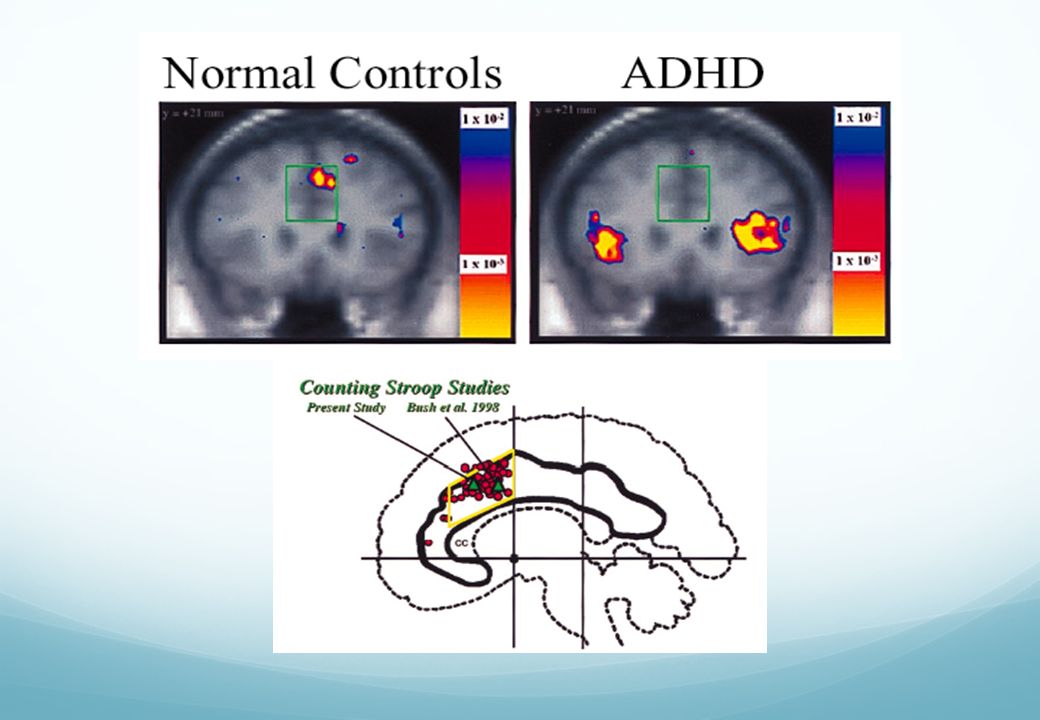
I/ Le modèle classique Hypofonctionnement du cortex frontal
= inhibition de l’action (Barkley) Imagerie fonctionnelle dans des protocoles d’inhibition (go-no-go, stop signal, flanker, Stroop….) Hypofonctionnement du cortex frontal Notion de dysfonctionnement exécutif
 Imagerie fonctionnelle dans des protocoles d’inhibition (go-no-go, stop signal, flanker, Stroop….) Hypofonctionnement du cortex frontal. Notion de dysfonctionnement exécutif.")
8
Inhibition comportementale
Contrôle moteur, fluence, syntaxe Mémoire de travail (non verbale) Internalisation de la parole Autorégulation de l’affect/ motivation/éveil reconstitution Barkley, R.A. (1997; 2007)
 Internalisation de la parole. Autorégulation de l’affect/ motivation/éveil. reconstitution. Barkley, R.A. (1997; 2007)")
10
Substrat cérébral du trouble attentionnel
Travaux de Posner et coll. Le triple système de l'attention selon Posner : alerte, orientation, contrôle exécutif orienting alerting Executive (conflict)
")
11
Sillon intra-pariétal/ Lobule pariétal supérieur Frontal Eye Field
Réseau fronto-pariétal dorsal (bilatéral) : attention endogène; génération d’un « set attentionnel » applicable lors du traitement du stimulus dans une tâche donnée Sillon intra-pariétal/ Lobule pariétal supérieur Frontal Eye Field Cortex visuel Jonction temporo-pariétale (lobule pariétal inférieur, gyrus temporal supérieur Cortex frontal ventral (gyrus frontal moyen et inférieur) Réseau fronto-pariétal ventral (fortement latéralisé à droite) : attention exogène; détection de stimuli comportementalement pertinents. Système d’alerte pour le système dorsal. Corbetta et al., 2002
 : attention endogène; génération d’un « set attentionnel » applicable lors du traitement du stimulus dans une tâche donnée. Sillon intra-pariétal/ Lobule pariétal supérieur. Frontal Eye Field. Cortex visuel. Jonction temporo-pariétale. (lobule pariétal inférieur, gyrus temporal supérieur. Cortex frontal ventral. (gyrus frontal moyen et inférieur) Réseau fronto-pariétal ventral (fortement latéralisé à droite) : attention exogène; détection de stimuli comportementalement pertinents. Système d’alerte pour le système dorsal. Corbetta et al.,")
12
Bilatéral et symétrique
« Dorsal attentional network » : orientation spatiale « Ventral attentional network » : détection événements motivants Fortement latéralisé à droite Thiebaut de Schotten, 2011

13
The SLF II connects the parietal component of the VAN to the prefrontal component of the DAN

14
Cortex pré-frontal dorso-latéral
Cingulaire dorsal antérieur Cortex pariétal Cortex pré-frontal ventro-latéral Striatum : caudé et putamen cervelet Les principales régions cérébrales dysfonctionnelles dans le TDAH (méta-analyse)
")
17
Twenty-one data sets were included for inhibition (287 patients with ADHD and 320 control subjects), and 13 data sets were included for attention (171 patients with ADHD and 178 control subjects). . Inhibition tasks and attention tasks. A, All inhibition tasks together. Regions of decreased (red and orange) and increased (blue) activation in patients with attention-deficit/hyperactivity disorder compared with healthy controls. Decreased activation in patients with attention-deficit/hyperactivity disorder relative to healthy controls is shown in the right inferior prefrontal cortex (IFC) extending into the insula, in a cluster comprising the supplementary motor area (SMA) and the cognitive division of anterior cingulate cortex (ACC), in the left caudate extending into the putamen and insula, and in the right mid-thalamus. B, Attention tasks. Decreased activation in patients with attention-deficit/hyperactivity disorder is shown in the right dorsolateral prefrontal cortex (DLPFC), in the left putamen and globus pallidus, in the right posterior thalamus (pulvinar) and caudate tail extending into the posterior insula, in the right inferior parietal lobe, and in the precuneus and superior temporal lobe. Increased activation in patients with attention-deficit/hyperactivity disorder relative to healthy controls was seen in the left cuneus and in the right cerebellum.
 and increased (blue) activation in patients with attention-deficit/hyperactivity disorder compared with healthy controls. Decreased activation in patients with attention-deficit/hyperactivity disorder relative to healthy controls is shown in the right inferior prefrontal cortex (IFC) extending into the insula, in a cluster comprising the supplementary motor area (SMA) and the cognitive division of anterior cingulate cortex (ACC), in the left caudate extending into the putamen and insula, and in the right mid-thalamus. B, Attention tasks. Decreased activation in patients with attention-deficit/hyperactivity disorder is shown in the right dorsolateral prefrontal cortex (DLPFC), in the left putamen and globus pallidus, in the right posterior thalamus (pulvinar) and caudate tail extending into the posterior insula, in the right inferior parietal lobe, and in the precuneus and superior temporal lobe. Increased activation in patients with attention-deficit/hyperactivity disorder relative to healthy controls was seen in the left cuneus and in the right cerebellum.")
19
Méta-analyse de 7 études de la substance grise (143 témoins vs 114 patients TDAH) : seule significativité = putamen/pallidum droit
 : seule significativité = putamen/pallidum droit")
20
II/ Le modèle alternatif
= défaut des systèmes de la récompense (Sonuga-Barke) Imagerie fonctionnelle dans des protocoles de récompense Dysfonctionnement des circuits cortico-striés
 Imagerie fonctionnelle dans des protocoles de récompense. Dysfonctionnement des circuits cortico-striés.")
21
Système dopaminergique
Voie méso-limbique Voie méso-corticale Aire septale Aire tegmentale ventrale Système dopaminergique Voie nigro-striée

22
Noyau accumbens Chez l'animal : en activité lors de l'anticipation d'une récompense, l' évaluation de la magnitude d'une récompense Chez l'homme impliqué dans les conduites à risque et addictives

23
Le « système de la récompense »
Noyau accumbens activé lors d’une tâche de gambling en IRM fonctionnelle Le « système de la récompense »

24
Physiopathologie du TDAH (D’après Sonuga-Barke, 2002, 2003)
")
25
Dopamine D 2/D3 receptor availability
Dopamine transporter availability

26
Haber, S.N. (2003) The primate basal ganglia: parallel and integrative networks. J. Chem. Neuroanat. 26, 317–330

27
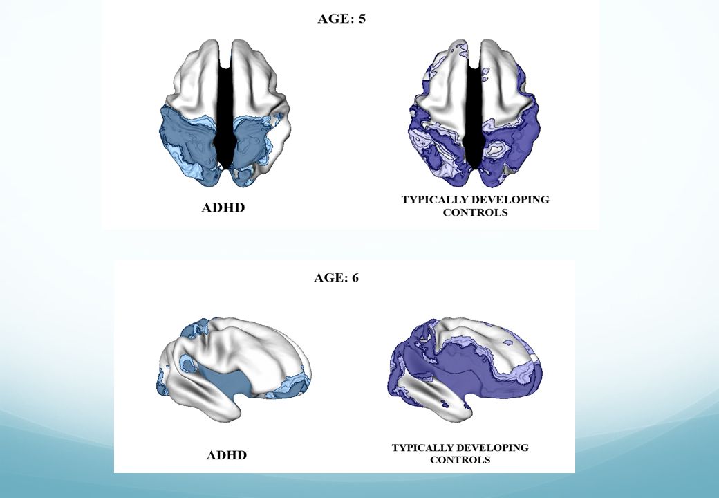
In the ventral striatal surfaces, there was a diagnostic difference in developmental trajectories (t = 5.6, p < .0001). Here, the typically developing group showed surface area expansion with age (estimated rate of increase of 0.54 mm2 per year, standard error [SE] 0.29 mm2 per year), whereas the ADHD group showed progressive contraction (decrease of 1.75 mm2 per year, SE 0.28 mm2 per year). The ADHD group also showed significant, fixed surface area reductions in dorsal striatal regions, which were detected in childhood at study entry and persisted into adolescence.
, whereas the ADHD group showed progressive contraction (decrease of 1.75 mm2 per year, SE 0.28 mm2 per year). The ADHD group also showed significant, fixed surface area reductions in dorsal striatal regions, which were detected in childhood at study entry and persisted into adolescence..")
29
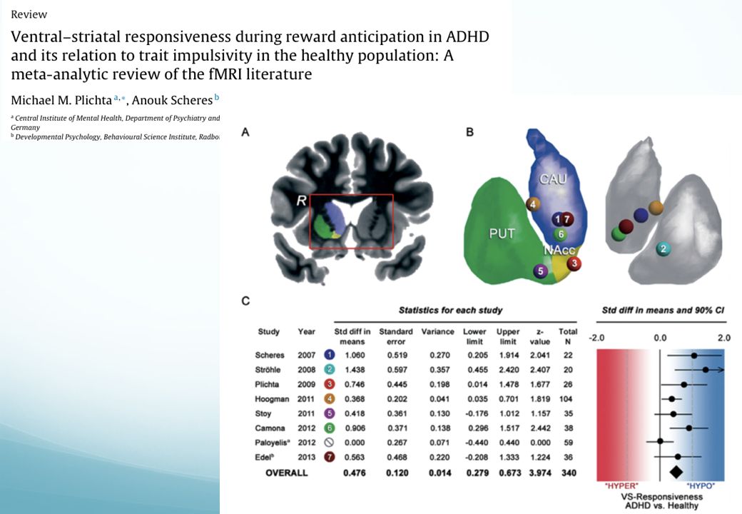
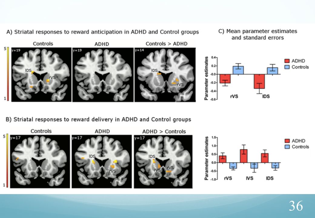
TDAH : hypoactivation du striatum ventral lors de l'anticipation de la récompense

31
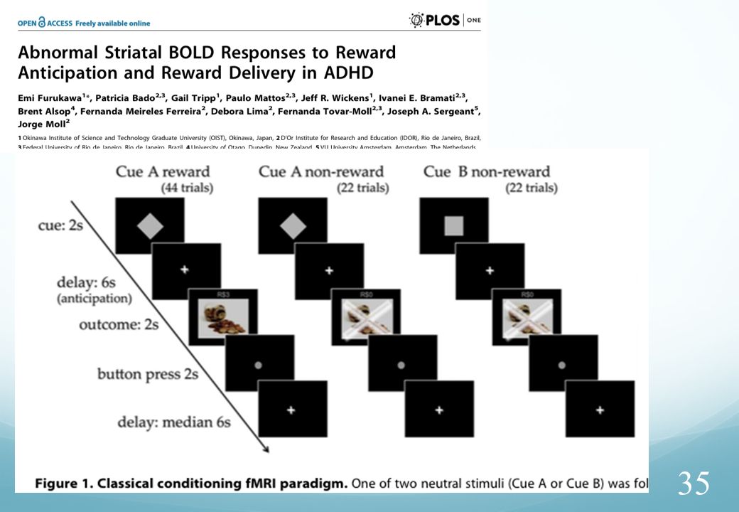
delayed rewards evoked hyperactivation in dorsal
ADHD : hyporesponsiveness of the ventral-striatal reward system for both immediate and delayed rewards delayed rewards evoked hyperactivation in dorsal caudate nucleus and amygdala of ADHD patients.

32
Sustained attention Reward
Areas of hypoactivation in CD children compared to ADHD and controls Sustained attention (Hippoc.+insula) Reward (ventro-lateral orbital)
 Reward. (ventro-lateral orbital)")
33
Comparaison adulte/enfant :
tâche d’attention soutenue : hypoactivation fronto-pariéto-striatale (comme chez l’enfant) Tâche récompensée : hypoactivation ventro-médiane, mais seulement si trouble des conduites comorbide Rewarded CPT 1£ pour 3 bonnes réponses (hits)
 Tâche récompensée : hypoactivation ventro-médiane, mais seulement si trouble des conduites comorbide. Rewarded CPT. 1£ pour 3 bonnes réponses (hits)")
34
Effet de la cible : sous-activation fronto-striatale
Effet de la récompense : sous activation frontale antérieure gauche

37
Fonctions exécutives « froides » Fonctions exécutives « chaudes »
CPFDL CFI CCAd AMS COF latéral COFvm CCAv Caudé/putamen/pallidum Striatum ventral Amygdala Temporo-pariétal The review together with the new data therefore suggest that childhood and adult ADHD, as a group, are characterised by the impairment of several overlapping neural networks, affecting fronto-subcortical, fronto-cortical and fronto-cerebellar neural circuitries that mediate “cool” attention and cognitive control functions as well as fronto-temporo-limbic neural networks of affect and motivation control (see schematic Fig. 6). The “cool” neural circuitries furthermore are known to interact closely with the “hot” neural circuitries (Goel and Dolan, 2003 and Krain et al., 2006). The findings are therefore in line with previously suggested concepts of multiple neural system impairment in ADHD associated with the different motor, attention, cognitive control and motivational processes that are impaired in the disorder (Makris et al., 2009 and Nigg and Casey, 2005). Schematic representation of the MRI evidence for structural and functional brain abnormalities in children and adults with ADHD in overlapping neural networks that mediate “cool” cognitive-abstract and “hot” reward-associated executive and cognitive functions. “Cool” cognitive networks of dysfunction in ADHD include inferior, dorsolateral and medial fronto-striatal, fronto-parieto-temporal and fronto-cerebellar regions and networks that mediate functions such as motor response and interference inhibition, cognitive flexibility, temporal foresight, selective and sustained attention, working memory, motor and timing processes. “Hot” EF network dysfunctions in ADHD patients have been observed in the context of temporal discounting, reward processing and reward anticipation. IFG = inferior frontal gyrus; OFC = orbitofrontal cortex; DLPFC = dorsolateral prefrontal cortex; vmOFC = ventromedial orbitofrontal cortex; d/vACC = dorsal/ventral ACC cortex; SMA = Supplementary Motor Area. Temporal supérieur Cervelet
. The cool neural circuitries furthermore are known to interact closely with the hot neural circuitries (Goel and Dolan, 2003 and Krain et al., 2006). The findings are therefore in line with previously suggested concepts of multiple neural system impairment in ADHD associated with the different motor, attention, cognitive control and motivational processes that are impaired in the disorder (Makris et al., 2009 and Nigg and Casey, 2005). Schematic representation of the MRI evidence for structural and functional brain abnormalities in children and adults with ADHD in overlapping neural networks that mediate cool cognitive-abstract and hot reward-associated executive and cognitive functions. Cool cognitive networks of dysfunction in ADHD include inferior, dorsolateral and medial fronto-striatal, fronto-parieto-temporal and fronto-cerebellar regions and networks that mediate functions such as motor response and interference inhibition, cognitive flexibility, temporal foresight, selective and sustained attention, working memory, motor and timing processes. Hot EF network dysfunctions in ADHD patients have been observed in the context of temporal discounting, reward processing and reward anticipation. IFG = inferior frontal gyrus; OFC = orbitofrontal cortex; DLPFC = dorsolateral prefrontal cortex; vmOFC = ventromedial orbitofrontal cortex; d/vACC = dorsal/ventral ACC cortex; SMA = Supplementary Motor Area. Temporal supérieur. Cervelet.")
38
Conclusion La dysfonction des systèmes exécutifs « froids », sous-tendus par le cortex frontal latéral, reste une explication valide de la limitation des capacités attentionnelles, en particulier l’attention soutenue L’impulsivité et l’aversion au délai (« hot-executive »), sous- tendus par les circuits à origine orbito-frontale, seraient plutôt liés à un défaut d’ajustement des systèmes de la récompense, en particulier lors de comorbidité avec des troubles des conduites Il y a de forts arguments, en particulier anatomiques, pour présumer que le primum movens est une dysfonction au niveau des circuits de la récompense et que les autres systèmes sont secondairement dysfonctionnels
, sous- tendus par les circuits à origine orbito-frontale, seraient plutôt liés à un défaut d’ajustement des systèmes de la récompense, en particulier lors de comorbidité avec des troubles des conduites. Il y a de forts arguments, en particulier anatomiques, pour présumer que le primum movens est une dysfonction au niveau des circuits de la récompense et que les autres systèmes sont secondairement dysfonctionnels.")
39
III/ défaut de connectivité et mécanismes du trouble de l’attention.
= défaut de connectivité de repos (« default mode) + tractographie : étude des circuits cortico-sous-corticaux Dysfonctionnement des connexions cortico- corticales et cortico-sous-corticales
 + tractographie : étude des circuits cortico-sous-corticaux. Dysfonctionnement des connexions cortico- corticales et cortico-sous-corticales.")
40
État de repos (resting state) enregistré en IRMf BOLD chez 20 adultes TDAH et 20 témoins appariés : anticorrélation entre DMN (précuneus et ventromedial prefrontal cortex) et cingulaire antérieur. La moindre activité chez les TDAH suggère un défaut de connectivité entre ces deux structures et un défaut de suppression de l'activité spontanée (non dirigée vers un but) (Castellanos et al., Biol. Psych., 2008).
.")
41
Tâche orientée vers un but
repos Tâche orientée vers un but Attention prêtée à la tâche 100 110 120 130 140 150 160 180 190 200 210 220 230 240 250 260 270 280 290 50 60 70 80 90 300 Seuil d’interférence TEMPS (Sec) Activité extrospective Activité introspective Chez l’hyperactif, une activité mentale spontanée excessive au repos empêcherait la cessation de ces fluctuations qui continueraient après le début de l’action, parasitant ainsi son bon déroulement, ce qui se traduirait par l’observation des troubles de l’attention
 Activité extrospective. Activité introspective. Chez l’hyperactif, une activité mentale spontanée excessive au repos empêcherait la cessation de ces fluctuations qui continueraient après le début de l’action, parasitant ainsi son bon déroulement, ce qui se traduirait par l’observation des troubles de l’attention.")
42
Psychiatry Res: Neuroimage. 2012;202:150-154
Compromised white matter integrity in attention deficit-hyperactivity disorder (ADHD). Regions of significant differences between adolescents with ADHD and controls shown in coronal, axial and sagittal views from the tract-based spatial statistics analysis. The white matter skeleton used in this analysis is displayed in yellow. Regions in which children with ADHD had higher fractional anisotropy (FA) are shown in red. Regions in which children with ADHD had higher axial diffusivity (AD) values than controls are shown in light blue. Group differences were “thickened” for visualization purposes, shown in red and blue for FA and AD respectively (ie, lighter colors represent the actual skeleton and the darker colors are the areas that were “thickened”). The bottom right panel of the figure shows the frontostriatal mask used in the analysis. Reproduced from ref 109: Tamm L, Bamea-Goralv N, Reiss AL. Diffusion tensor imaging reveals white matter abnormalities in Attention-Deficit/Hyperactivity Disorder. Psychiatry Res: Neuroimage. 2012;202: Copyright © Elsevier 2012 DTI : Regions of significant differences between adolescents with ADHD and controls
. Regions of significant differences between adolescents with ADHD and controls shown in coronal, axial and sagittal views from the tract-based spatial statistics analysis. The white matter skeleton used in this analysis is displayed in yellow. Regions in which children with ADHD had higher fractional anisotropy (FA) are shown in red. Regions in which children with ADHD had higher axial diffusivity (AD) values than controls are shown in light blue. Group differences were thickened for visualization purposes, shown in red and blue for FA and AD respectively (ie, lighter colors represent the actual skeleton and the darker colors are the areas that were thickened ). The bottom right panel of the figure shows the frontostriatal mask used in the analysis. Reproduced from ref 109: Tamm L, Bamea-Goralv N, Reiss AL. Diffusion tensor imaging reveals white matter abnormalities in Attention-Deficit/Hyperactivity Disorder. Psychiatry Res: Neuroimage. 2012;202: Copyright © Elsevier DTI : Regions of significant differences between adolescents with ADHD and controls.")
43
Corrélation entre défaut de connectivité dans divers faisceaux d’association et nombre d’erreurs d’omission au CPT2 ADHD, combined (n=39) ADHD, inattentive (n=26) Department of Psychiatry (S-BH, AZ, AF), Melbourne Neuropsy- chiatry Centre, University of Melbourne and Melbourne Health; and Florey Institute of Neuroscience and Mental Health (S-BH), Parkville, Victoria, Australia; Division of Child and Adolescent Psychiatry (S-BH, SP, M-HP, M-SS, B-NK, S-CC, J-WK), Department of Psychiatry, College of Medicine, Seoul National University, Seoul, Republic of Korea;
 ADHD, inattentive (n=26) Department of Psychiatry (S-BH, AZ, AF), Melbourne Neuropsy- chiatry Centre, University of Melbourne and Melbourne Health; and Florey Institute of Neuroscience and Mental Health (S-BH), Parkville, Victoria, Australia; Division of Child and Adolescent Psychiatry (S-BH, SP, M-HP, M-SS, B-NK, S-CC, J-WK), Department of Psychiatry, College of Medicine, Seoul National University, Seoul, Republic of Korea;")
44
Méta-analyse « ALE » de 15 études DTI publiées à 2011:173 ADHD patients and 169 healthy control subjects Diminution d’anisotropie -faisceau long. sup - f. cortico-spinal connexions fronto-striées Cervelet gauche

45
③ ② c ④ ① ⑤ Corrélé à l’impulsivité Proportionnel aux mesures d’attention TDAH :schématisation des principaux faisceaux d’association issus du cortex frontal tels qu’ils peuvent être individualisés en tractographie IRM. anomalie d’anisotropie en tractographie proportionnelle à diverses mesures des fonctions exécutives (mémoire de travail, attention soutenue, flexibilité...) 4, 5 : fronto-strié ventro-latéral et orbito-caudé : corrélés au déficit attentionnel 1 : unciné : corrélé à l’impulsivité D’après Chang et al. (2012); Wu et al. (2012) Figure : schématisation des principaux faisceaux d’association issus du cortex frontal tels qu’ils peuvent être individualisés en tractographie IRM. 1: faisceau unciné (unissant le cortex fronto-orbitaire et la région de l’amygadala, en rouge). 2- faisceau préfronto-caudé médian (région cingulaire — partie antérieure du noyau caudé); 3- préfrontal dorso-latéral — caudé latéral); 4- pré-frontal ventro-latéral — caudé antérieur); 5- orbito-frontal — caudé inférieur (noyau accumbens)
 4, 5 : fronto-strié ventro-latéral et orbito-caudé : corrélés au déficit attentionnel. 1 : unciné : corrélé à l’impulsivité. D’après Chang et al. (2012); Wu et al. (2012) Figure : schématisation des principaux faisceaux d’association issus du cortex frontal tels qu’ils peuvent être individualisés en tractographie IRM. 1: faisceau unciné (unissant le cortex fronto-orbitaire et la région de l’amygadala, en rouge). 2- faisceau préfronto-caudé médian (région cingulaire — partie antérieure du noyau caudé); 3- préfrontal dorso-latéral — caudé latéral); 4- pré-frontal ventro-latéral — caudé antérieur); 5- orbito-frontal — caudé inférieur (noyau accumbens)")
46
Conclusion (2) Les méthodes de tractographie ont récemment été largement utilisées dans le TDAH, pour évaluer la thèse d’un défaut de connectivité. Un défaut de connectivité de divers circuits cortico-corticaux et cortico- sous-corticaux pourrait rendre compte de ces déficits, et aussi de l’activité mentale spontanée matéralisée par l’activité de repos dans le circuit « default-mode ». De nombreux faisceaux de substance blanche ont été analysés comme anormalement constitués ou organisés dans le TDAH (enfant et adulte), à divers niveaux : cortico-cortical, cortico-strié, cortico- cérébelleux. Les travaux les plus récents pointent vers un défaut multifocal de connectivité avec des variations individuelles pouvant correspondre à la présence des différents symptômes : inattention, agitation- impulsivité, signes moteurs, troubles des conduites
, à divers niveaux : cortico-cortical, cortico-strié, cortico- cérébelleux. Les travaux les plus récents pointent vers un défaut multifocal de connectivité avec des variations individuelles pouvant correspondre à la présence des différents symptômes : inattention, agitation- impulsivité, signes moteurs, troubles des conduites.")
Présentations similaires