Télécharger la présentation
La présentation est en train de télécharger. S'il vous plaît, attendez

1
Gelures Emmanuel Cauchy MD IFREMMONT Hôpital de Chamonix
Courmayeur Novembre 2008 Firstly, I would like to thank you for the invitation. I’m very happy to present here the results of 5 years of study concerning 20 years of frostbite injuries managment in Chamonix Hospital on about 1800 cases of frostbite injuries. I will try to synthesize the lecture in order to obtain a usefull algorithm in the aim of giving the possibilty to any medical doctor or rescuer the means of curing or dispatching the patients in the correct way I thank as well, Bernard Marsigny, unfortunately not here today for his help and Dr Foray for his remarkable archievements in frostbite documentation.

4
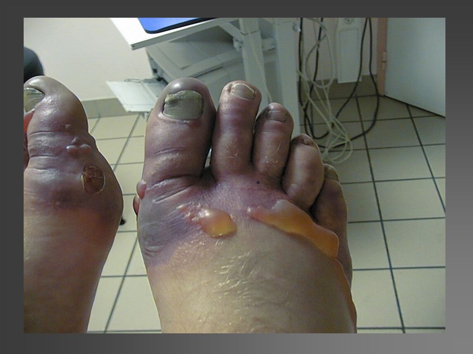
Severe frostbite of the feet on a young alpinist D+36h
For example, this very good young mountaineer was stuck for six days on the north face of the Matternhorn whilst attempting a solo route. This picture shows us the state of his feet thirty six hours after his rescue. We can observe the presence of clear blisters and an absence of necrotic lesions. If we refer to the traditional classification, we can evaluate this lesion in Grade II A, which means a good pronogsis without amputation.

5
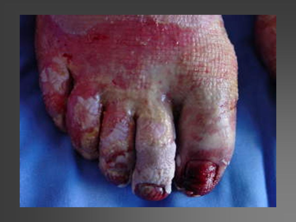
Severe frostbite of the feet on a young alpinist D+36h
For example, this very good young mountaineer was stuck for six days on the north face of the Matternhorn whilst attempting a solo route. This picture shows us the state of his feet thirty six hours after his rescue. We can observe the presence of clear blisters and an absence of necrotic lesions. If we refer to the traditional classification, we can evaluate this lesion in Grade II A, which means a good pronogsis without amputation.

6
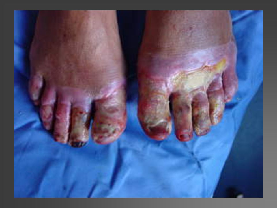
Severe frostbite of the feet on a young alpinist D+36h
For example, this very good young mountaineer was stuck for six days on the north face of the Matternhorn whilst attempting a solo route. This picture shows us the state of his feet thirty six hours after his rescue. We can observe the presence of clear blisters and an absence of necrotic lesions. If we refer to the traditional classification, we can evaluate this lesion in Grade II A, which means a good pronogsis without amputation.

7
Rappel sur la gelure Définition Lésion localisée résultante d’une exposition prolongée à une température inférieure à 0°C Contexte Urbain +++ Terrain d’aventure Before starting, I would like to make a quick reminder of the definition The frostbite injury is a localized lesion, normally a direct result of the cold and exposure for a considerable length of time at a temperature below 0°Celcius It most often occurs on the hands or feet, but more rarely on the cheeks, ears, nose, elbow, patella, buttocks or foreskin.

8
Mécanisme Homéothermie Deux compartiments, l’écorce et le noyau …
Vasoconstriction périphérique Shunts artério-veineux Le noyau stable Protection des organes nobles (reins, foie, cerveau, cœur)
")
9
Le lapin Objectif prioritaire « lutter contre la phase de nécrose progressive »

10
Physiopathologie Phase primaire : refroidissement et action du gel
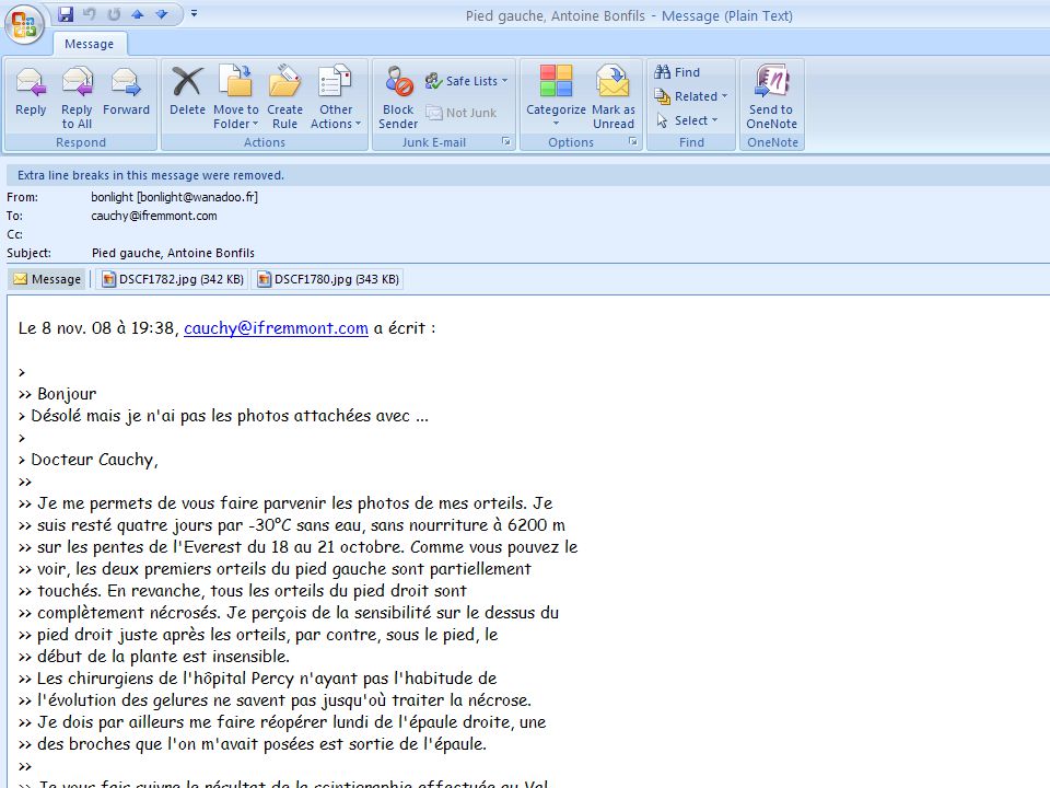
Vasoconstriction périphérique Fermeture des sphincters précapillaires et ouverture des shunts artério-veineux (poignets et chevilles) Mort cellulaire par agression mécanique (recristallisation) et déshydratation cellulaire Phase secondaire : réchauffement et nécrose progressive Syndrome d'ischémie-reperfusion [3] caractérisé par la libération de substances vaso-actives aboutissant en quelques heures à un arrêt complet de la microcirculation. Phase tardive : les lésions définitives C'est une phase lente et progressive qui peut prendre plusieurs semaines (J7-J45), les tissus revascularisés se réorganisent et se cicatrisent alors que les tissus dévitalisés évoluent lentement vers la gangrène sèche irréversibles. CLINIQUE et PHYSIOPATHOLOGIE Phase primaire : refroidissement et action du gel A cette phase, la clinique est pauvre, essentiellement marquée par un aspect livide et froid des tissus atteints. L'anesthésie induite par le froid rend cette phase indolore [Photo 1]. L'organisme soumis au froid présente une vasoconstriction périphérique dont l'importance dépend de l'intensité du froid et du tonus vasomoteur de l'individu. Cette vasoconstriction artérielle et veineuse avec détournement sanguin par les anastomoses artério-veineuses et fermeture des sphincters précapillaires entraîne une diminution du gradient de perfusion capillaire et l'apparition de phénomènes locaux de stagnation, d'hyperviscosité, d'hypoxie et d'acidose. La cause de la mort cellulaire varie en fonction de la vitesse d'installation des lésions. Souvent consécutive à l'agression mécanique des cristaux extracellulaires, sa cause peut également être intracellulaire par l'aboutissement du phénomène de déshydratation et de recristallisation qui peut être limité par le réchauffement rapide en faisant fondre les cristaux avant qu'ils n'augmentent de taille [2]. Phase secondaire : réchauffement et nécrose progressive Sur le plan clinique, c'est la phase la plus riche (œdème, phlyctènes, nécrose), elle débute dès la phase de réchauffement. Elle est caractérisée par la démarcation de la lésion initiale, grise, cyanosée, peu sensible au toucher. Quand la gelure atteint les extrémités, elle évolue de manière centripète remontant de la pulpe vers la racine des membres [photo 2]. Cette lésion initiale persiste 12 à 24 heures avant l'apparition des phlyctènes. Absentes lors des gelures superficielles, les phlyctènes peuvent être hématiques, séro-hématiques voire hémorragiques quand les gelures sont plus graves [photo 3]. Elles sont parfois volumineuses et compressives. Elle peuvent apparaître dès la douzième heure et persister plusieurs jours (J1-J7) . En l'absence d'excision chirurgicale, elles finissent par se rompre spontanément. Sur le plan physiopathologique, c'est le début de la nécrose secondaire progressive avec syndrome d'ischémie-reperfusion [3] caractérisé par la libération de substances vaso-actives aboutissant en quelques heures à un arrêt complet de la microcirculation. Phase tardive : les lésions définitives C'est une phase lente et progressive qui peut prendre plusieurs semaines (J7-J45), les tissus revascularisés se réorganisent et se cicatrisent alors que les tissus dévitalisés évoluent lentement vers la gangrène sèche se démarquant du reste par un sillon d'élimination [photo 4]. Les lésions sont alors irréversibles et si le traitement n'est débuté qu'à ce stade, les résultats sont décevants.
 Mort cellulaire par agression mécanique (recristallisation) et déshydratation cellulaire. Phase secondaire : réchauffement et nécrose progressive. Syndrome d ischémie-reperfusion [3] caractérisé par la libération de substances vaso-actives aboutissant en quelques heures à un arrêt complet de la microcirculation. Phase tardive : les lésions définitives. C est une phase lente et progressive qui peut prendre plusieurs semaines (J7-J45), les tissus revascularisés se réorganisent et se cicatrisent alors que les tissus dévitalisés évoluent lentement vers la gangrène sèche irréversibles. CLINIQUE et PHYSIOPATHOLOGIE. Phase primaire : refroidissement et action du gel. A cette phase, la clinique est pauvre, essentiellement marquée par un aspect livide et froid des tissus atteints. L anesthésie induite par le froid rend cette phase indolore [Photo 1]. L organisme soumis au froid présente une vasoconstriction périphérique dont l importance dépend de l intensité du froid et du tonus vasomoteur de l individu. Cette vasoconstriction artérielle et veineuse avec détournement sanguin par les anastomoses artério-veineuses et fermeture des sphincters précapillaires entraîne une diminution du gradient de perfusion capillaire et l apparition de phénomènes locaux de stagnation, d hyperviscosité, d hypoxie et d acidose. La cause de la mort cellulaire varie en fonction de la vitesse d installation des lésions. Souvent consécutive à l agression mécanique des cristaux extracellulaires, sa cause peut également être intracellulaire par l aboutissement du phénomène de déshydratation et de recristallisation qui peut être limité par le réchauffement rapide en faisant fondre les cristaux avant qu ils n augmentent de taille [2]. Phase secondaire : réchauffement et nécrose progressive. Sur le plan clinique, c est la phase la plus riche (œdème, phlyctènes, nécrose), elle débute dès la phase de réchauffement. Elle est caractérisée par la démarcation de la lésion initiale, grise, cyanosée, peu sensible au toucher. Quand la gelure atteint les extrémités, elle évolue de manière centripète remontant de la pulpe vers la racine des membres [photo 2]. Cette lésion initiale persiste 12 à 24 heures avant l apparition des phlyctènes. Absentes lors des gelures superficielles, les phlyctènes peuvent être hématiques, séro-hématiques voire hémorragiques quand les gelures sont plus graves [photo 3]. Elles sont parfois volumineuses et compressives. Elle peuvent apparaître dès la douzième heure et persister plusieurs jours (J1-J7) . En l absence d excision chirurgicale, elles finissent par se rompre spontanément. Sur le plan physiopathologique, c est le début de la nécrose secondaire progressive avec syndrome d ischémie-reperfusion [3] caractérisé par la libération de substances vaso-actives aboutissant en quelques heures à un arrêt complet de la microcirculation. Phase tardive : les lésions définitives. C est une phase lente et progressive qui peut prendre plusieurs semaines (J7-J45), les tissus revascularisés se réorganisent et se cicatrisent alors que les tissus dévitalisés évoluent lentement vers la gangrène sèche se démarquant du reste par un sillon d élimination [photo 4]. Les lésions sont alors irréversibles et si le traitement n est débuté qu à ce stade, les résultats sont décevants.")
11
Clinique 4 périodes successives Période de gel des tissus
Période de réchauffement Période de nécrose progressive tardive Période de momification

12
Période de gel des tissus
Aspect congelé Rigide voire dure Insensible Centripète In fact, serious frostbite always evolves in the same way. First, the frozen period is characterized by the cold, white, rigid aspect and insensitive of the extremities. It’s before the rewarming time

13
Période de réchauffement (J0)
Lésion initiale Aspect gris ou cyanosé Hypo ou anesthésie Centripète Insensible Température pulpaire --- At day 0, just after rewarming, there are no blisters or necrotic areas but just a pallid, cold, insensitive and sometimes cyanotic lesions.

14
Période de nécrose progressive tardive (J1-J3)
Apparition des phlyctènes Séro hématique ou hémorragiques Volumineuses Compressives A exciser (J3-J5) 24 to 36 hours later, blisters appear if the frostbite is sufficiently serious which could be voluminous, compressive and sometimes hemorrhagic. Physiologically, this second phase is characterized by a secondary belated necrosis. It is a phenomenon of ischemia reperfusion which leads to an extended area of the lesion.
 24 to 36 hours later, blisters appear if the frostbite is sufficiently serious which could be voluminous, compressive and sometimes hemorrhagic. Physiologically, this second phase is characterized by a secondary belated necrosis. It is a phenomenon of ischemia reperfusion which leads to an extended area of the lesion.")
15
Période de nécrose progressive tardive (J1-J3)
Apparition des phlyctènes Séro hématique ou hémorragiques Volumineuses Compressives A exciser (J3-J5) 24 to 36 hours later, blisters appear if the frostbite is sufficiently serious which could be voluminous, compressive and sometimes hemorrhagic. Physiologically, this second phase is characterized by a secondary belated necrosis. It is a phenomenon of ischemia reperfusion which leads to an extended area of the lesion.
 24 to 36 hours later, blisters appear if the frostbite is sufficiently serious which could be voluminous, compressive and sometimes hemorrhagic. Physiologically, this second phase is characterized by a secondary belated necrosis. It is a phenomenon of ischemia reperfusion which leads to an extended area of the lesion.")
16
Période de nécrose progressive tardive (J1-J3)
Apparition des phlyctènes Séro hématique ou hémorragiques Volumineuses Compressives A exciser (J3-J5) 24 to 36 hours later, blisters appear if the frostbite is sufficiently serious which could be voluminous, compressive and sometimes hemorrhagic. Physiologically, this second phase is characterized by a secondary belated necrosis. It is a phenomenon of ischemia reperfusion which leads to an extended area of the lesion.
 24 to 36 hours later, blisters appear if the frostbite is sufficiently serious which could be voluminous, compressive and sometimes hemorrhagic. Physiologically, this second phase is characterized by a secondary belated necrosis. It is a phenomenon of ischemia reperfusion which leads to an extended area of the lesion.")
17
Période de momification (….J45…)
Scarification Momification During the days which follow the secondary necrosis period, we see the progressive apparition of necrotic areas. They dry out and lyophilize. This period is commonly called scarification or mummification. The dividing line between dead and healthy skin becomes evident after three weeks of evolution : This is a dry gangrene. This is the evolutionary phase ( 3 weeks to one month later) when the surgeon decides to amputate.
 when the surgeon decides to amputate.")
18
Comment évaluer la gravité ?
Risque d’amputation? Type de traitement ? Durée du traitement ? Hospitalisation ? Séquelles fonctionnelles ? WHAT KIND OF INFORMATION CAN YOU USE TO ANSWER TO THESE QUESTIONS: Firstly : What kind of treatment do I have to propose ? Secondly : does he need any hospitalisation ? thirdly: What is the prognosis, does he need amputation and at what level ?

19
Prise en charge initiale
Protocole de réchauffement rapide Perfusion de 400 mg de chlorhydrate de buflomédil Aspirine 250 mg IV bolus Évaluation du niveau d’extension Stade 1 Stade 2 Stade 3 Stade 4

20
Classification Clinique et Algorythme

21
Stade 1 Here is an example of distal frostbite Grade I just after rapid rewarming. The initial lesion has disappeared. There is only an erythema remaining. The treatment will be ambulatory by aspirin and vasodilator. No risk of amputation.

22
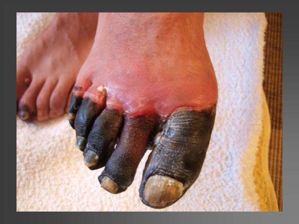
Stade 2 Here is frostbite Grade II
The initial lesion remains after rapid rewarming but stays limited to the distal phalanx. The treatment can be identical to Grade I. there will also be bandages for the blisters for two to three weeks. There is no risk of amputation, but there could be sores on the finger tips to excise.

23
Stade 3 Here is a frostbite Grade III
The initial lesion surpasses the distal phalanx, despite a rapid rewarming. Hospitalisation is recommended with intra-venous aspirin and vasodilator with daily bandages and antiseptic baths. A bone scanning should be performed from day 2 to determine the amputation areas to be expected.

24
Stade 4 Here is a frostbite at Grade IV
The patient must be hospitalized in intensive care. The level of amputation determined by the bone scan is substantial. It is sometimes neccessary to amputate urgently because there is a risk of this frostbite becoming infected and resulting in septicemia.

25
Risque d’amputation (95% CI )
Extension Risque d’amputation (95% CI ) Main 5 : carpe / tarse 100 % 4 : métacarpe / métatarse 3 : phalange proximale 83 % 2 : phalange intermédiaire 39 % 1 : phalange distale 1 % Pied 98 % 60 % 23 % 0 % et 67 % 31 % Using these two new criteria, we propose new early classification at two different times. At day 0, it can put into pratice at a distance and allows sufficiently precise information about the degree of severity and the risk of amputation. At day 2, it is made after an analysis of the results of the bone scan and gives a reliable and precise level of the amputations to expect. This classification is published in Wilderness and Environmental Medicine Journal. New classification of frostbite injuries of the extremities ( Wild Env J Med – 2001 ) Table 2 : New classification of the severity of frostbite injuries
 Main. 5 : carpe / tarse. 100 % 4 : métacarpe / métatarse. 3 : phalange proximale. 83 % 2 : phalange intermédiaire. 39 % 1 : phalange distale. 1 % Pied. 98 % 60 % 23 % 0 % et. 67 % 31 % Using these two new criteria, we propose new early classification at two different times. At day 0, it can put into pratice at a distance and allows sufficiently precise information about the degree of severity and the risk of amputation. At day 2, it is made after an analysis of the results of the bone scan and gives a reliable and precise level of the amputations to expect. This classification is published in Wilderness and Environmental Medicine Journal. New classification of frostbite injuries of the extremities ( Wild Env J Med – 2001 ) Table 2 : New classification of the severity of frostbite injuries.")
26
Scinti Osseuse au Tc 99m Seul examen complémentaire validé sur une grande série Non invasif Pronostic à J2 Médico-légal This picture illustrates the correlation between this examination and the final outcome. Whats more, this examination is non-invasive and can eventually be used as evidence in a medico-legal case. The Journal of Hand Surgery American (Sept 2000) European Journal of Nuclear Medicine (Mai 2000)
 European Journal of Nuclear Medicine (Mai 2000)")
27
Stade I Stade II Stade III Stade IV Traitement Gelures
Traitement Gelures Stade I Stade II Stade III Stade IV Stade d’extension après réchauffement rapide Pas de lésion initiale La dernière phalange Phalange intermédiaire Carpe / tarse Scintigraphie Osseuse à J2 Inutile Hypofixation Défect isotope sur le carpe / tarse Phlyctènes Pas de phlyctène Phlyctène séreuse Phlyctène hémorragique carpe / tarse Pronostic Pas d’amputation Pas de séquelle Amputation Tissulaire (pulpe) Séquelles Phanère Amputation osseuse distale Séquelles Fonctionnelles Amputation des membres +/- Atteinte systémique +/- Sepsis Séquelles fonctionnelles majeures Using these two new criteria, we propose new early classification at two different times. At day 0, it can put into pratice at a distance and allows sufficiently precise information about the degree of severity and the risk of amputation. At day 2, it is made after an analysis of the results of the bone scan and gives a reliable and precise level of the amputations to expect. This classification is published in Wilderness and Environmental Medicine Journal. New classification of frostbite injuries of the extremities ( Wild Env J Med – 2001 ) Table 2 : New classification of the severity of frostbite injuries
 Séquelles Phanère. Amputation osseuse distale. Séquelles Fonctionnelles. Amputation des membres. +/- Atteinte systémique. +/- Sepsis. Séquelles fonctionnelles majeures. Using these two new criteria, we propose new early classification at two different times. At day 0, it can put into pratice at a distance and allows sufficiently precise information about the degree of severity and the risk of amputation. At day 2, it is made after an analysis of the results of the bone scan and gives a reliable and precise level of the amputations to expect. This classification is published in Wilderness and Environmental Medicine Journal. New classification of frostbite injuries of the extremities ( Wild Env J Med – 2001 ) Table 2 : New classification of the severity of frostbite injuries.")
28
Stade I Stade II Stade III Stade IV
Stade I Stade II Stade III Stade IV Pas d’hospitalisation Pas de scintigraphie Hospitalisation 2 jours +/- Scintigraphie osseuse J2 Hospitalisation 8 jours Scintigraphie à J2 et J8 Hospitalisation USI Traitement oral 10 jours Aspirine + buflomédil Pas de traitement local Using these two new criteria, we propose new early classification at two different times. At day 0, it can put into pratice at a distance and allows sufficiently precise information about the degree of severity and the risk of amputation. At day 2, it is made after an analysis of the results of the bone scan and gives a reliable and precise level of the amputations to expect. This classification is published in Wilderness and Environmental Medicine Journal. Protocole randomisé Bufl. / Prost. / Thromb. Hydrocolloïde / +/- ABT +/- amputation en urgence Traitement IV aspirine + buflomédil Traitement local hydrocolloïde Protocole randomisé Bufl. / Prost. / Thromb. Hydrocolloïde +/- amputation New classification of frostbite injuries of the extremities ( Wild Env J Med – 2001 ) Table 2 : New classification of the severity of frostbite injuries
 Table 2 : New classification of the severity of frostbite injuries.")
29
Traitement Local

31
Pansements locaux hydro colloïdes

32
Traitement Aucun traitement validé actuellement !
Seule pratique évaluée sur la cellule animale réchauffement rapide Traitement empirique basé sur L’aspirine Les vasodilatateurs classiques (buflomédil, pentoxifilline) L’hémodilution Les AINS Les anticoagulants Parage des phlyctènes Antibiotiques en cas de surinfection Amputation tardive quand gangrène sèche Amputation rapide quand gangrène humide
 L’hémodilution. Les AINS. Les anticoagulants. Parage des phlyctènes. Antibiotiques en cas de surinfection. Amputation tardive quand gangrène sèche. Amputation rapide quand gangrène humide.")
33
Traitement Protocole expérimental aux hôpitaux du Mont-Blanc
A - Traitement classique Vasodilatateur classique , buflomedil (Fonzylane) Aspirine B – Traitement par prostacycline (prostanoïdes) Iloprost (Ilomedine) C – Traitement par thrombolitique r-TPase + Iloprost
 Aspirine. B – Traitement par prostacycline (prostanoïdes) Iloprost (Ilomedine) C – Traitement par thrombolitique. r-TPase + Iloprost.")
38
Traitement Chirurgical

39
Frostbite injury at the Everest July 2003

40
Frostbite injury at the Everest July 2003
Photo Dumontier

41
Frostbite injury at the Everest July 2003
Photo Dumontier

42
Frostbite injury at the Everest July 2003
Photo Dumontier

43
Frostbite injury at the Everest July 2003
Photo Dumontier J + 1 month + 2 month

44
Laser Dopler Imager The laser doppler imager is non invasive easy to do and rapid. It gives information on the surface micro-vascularisation of the skin which would be a reflection of the profound microvascularisation. The blue color corresponds to the frozen area on the thumb.

45
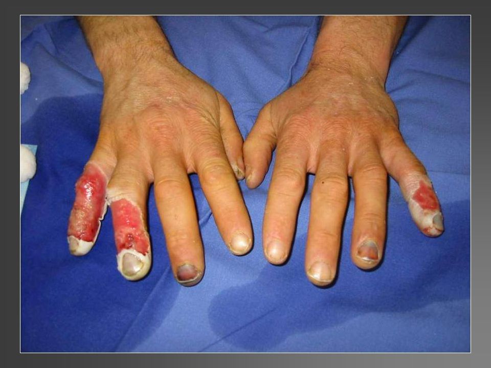
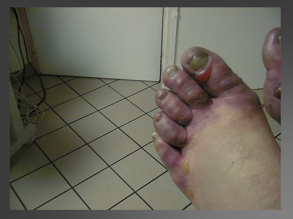
IRM This a picture of digitalized angiography with MRI, in the future we will try to validate this examination for the frostbite injury. It is able to give a very concise picture of the micro-vascularisation without an invasive effect contrary to the classical angiography. This patient presented a severe frostbite injury of the toes especially on the right.

46
3 éléments à retenir ! Lésion grave : en amont de la dernière phalange
Stade 3 et 4 : risque d’amputation à prendre en charge en urgence Nouveau protocole [prostacycline et thrombolytique] à appliquer en moins de 48h diminue fortement les risques d’amputation

Présentations similaires