Télécharger la présentation
La présentation est en train de télécharger. S'il vous plaît, attendez

1
HEPATITES CHRONIQUES Dr Belghazi

2
I-INTRODUCTION : Définition: Lésion hépatique nécrotique et inflammatoire persistante et progressive d’évolution chronique (plus de 6 mois) susceptible de conduire à la fibrose et à la cirrhose.
 susceptible de conduire à la fibrose et à la cirrhose.")
3
L’activité (évolutivité)
Activité clinique : existence de symptômes en particulier ictère Activité biochimique : degré d’élévation ALAT Activité histologique : intensité de la nécrose hépatocytaire et de l’infiltration inflammatoire

4
Rappel anatomique L’unité est le lobule centré par une veine centro-lobulaire (VCL) limité en périphérie par les espaces portes (EP)
 limité en périphérie par les espaces portes (EP)")
5
II-ANAPATH 1. Lésions élémentaires :
1.1 Infiltration inflammatoire : mononuclée (lymphocytes et plasmocytes) Elle prédomine dans l’espace porte et la région péri portale
 Elle prédomine dans l’espace porte et la région péri portale.")
6
II-ANAPATH 1. Lésions élémentaires :
1.2 Nécrose hépatocytaire : Elle est focale : Souvent péri portale Affectant hépatocytes situées en contact de l’espace porte = hépatocytes de la lame bordante nécrose parcellaire= piecemeal necrosis Parfois : nécrose pénètre dans le lobule intéressant les travées hépatocytaires et réunissant alors espace porte à la veine centrolobulaire = nécrose en pont Parfois affecte qq. Hépatocytes situées à l’intérieure même du lobule = nécrose intra lobulaire.

7
II-ANAPATH 1. Lésions élémentaires :
1.3 Fibrose : Débute dans région portale peri portale fibrose en pont (reliant éspace porte et veine centrolobulaire)
")
8
2. Evaluation semi -quantitative des lésions histologiques score de Métavir :
Activité absente Fibrose 1 Minime Portale 2 Modérée Portale + qq. Septas 3 4 Sévère Septale sans cirrhose cirrhose

10
III-ETUDE CLINIQUE : 1. Clinique : pauvre, asymptomatique, douleurs, asthénie, ictère, prurit 2. Bilan hépatique : Presque constamment anormaux Transaminases : 2 – 10 N, , ALAT>ASAT. PAL, BGGT, gamma globulines : normaux ou modérément élevés. 3. Morphologie : Echo peut être normale 4. Histologie. PBF: place de choix dans le diagnostic = lésions élémentaires.

11
IV-DIAGNOSTIC : 1. DIAGNOSTIC POSITIF 2.DIAGNOSTIC DIFFERENTIEL 3.DIAGNOSTIC ETIOLOGIQUE

12
IV-DIAGNOSTIC : 1. DC POSITIF
PBF Confirmation par histologie

13
2. DC DIFFERENTIEL 2.1 Eliminer hépatite aigue:

14
-Métaboliques : hémochromatose -Choléstatique : CBP, CSP
2. DC DIFFERENTIEL 2.2 Eliminer hépatopathie chronique : -Métaboliques : hémochromatose -Choléstatique : CBP, CSP -Hépatopathie alcoolique, -Vasculaires : SBC, foie cardiaque, maladie veino occlusive -Granulomatose hépatique :sarcoidose ,tuberculose -Infectieuses : bactérienne, parasitaire, virale à virus non hépatotrope -NASH .

15
V- DgC ETIOLOGIQUE 1-. HEPATITES VIRALES : B, C,et B-D a-Hépatite chronique B : -Problème de santé publique majeur, plus de 2000 millions ont été au contact du virus, 350 millions de porteurs chroniques. -Prévalence de l’Ag Hbs en Algérie: 2,18% (1998) -VHB humain fait partie de la famille Hepadnaviridae (virus hépatotropes à acide désoxyribonucléique [ADN])
 -VHB humain fait partie de la famille Hepadnaviridae (virus hépatotropes à acide désoxyribonucléique [ADN])")
16
-Transaminases (ALAT/ASAT) Marqueurs de l’activité de l’infection
Marqueur Signification 1- Ag HBs Infection 1’-AC anti-HBs Guérison (immunite définitive) 2- Ag HBc Hépatocyte 2’-AC HBc Stigmate d’infection (ancienne / récente) 3-Ag HBe Réplication 3’-AC anti-HBe Non réplication 4- DNA viral Réplication
 2- Ag HBc Hépatocyte 2’-AC HBc Stigmate d’infection (ancienne / récente) 3-Ag HBe Réplication 3’-AC anti-HBe Non réplication 4- DNA viral Réplication")
17
-Dc Souvent fortuit: - sérologie virale systématique: asthénie persistante, facteurs de risque (drogués IV, homosexuels, transfusés, portage familial), -complication révélatrice (ascite, hémorragie digestive, CHC) ALAT > à 2 fois NL pendant plus de 6 mois,
, -complication révélatrice (ascite, hémorragie digestive, CHC) ALAT > à 2 fois NL pendant plus de 6 mois,")
19
-Aspect en verre dépoli ou «ground glass» du cytoplasme des hépatocytes contenant l’Ag HBs. -TRT : Immuno-modulateurs : Interféron alpha- Interféron pegylé Antiviraux

20
b-Hépatite virale D: - co-infection : Ac anti-D type IgM et/ou Ag D sérique IgM anti-HBc + pour le VHB. Évolue habituellement vers la resolution -surinfection aiguë : Ac anti-D, de type IgM et totaux positifs, IgM anti-VHB négatifs pour le VHB. cirrhose

21
c- Hépatite virale C : - Sémiologie silencieuse ou non spécifique.
-Chronicité: 80 % environ. Cirrhose dans 10 à 20% 10 à 20 ans plus tard, CHC 1 à 4% par an. -Clinique: peu ou asymptomatique; asthénie, -ALAT modérément élevées (< à 5 x Nle), fluctuantes +.µ -Dg le plus souvent méconnue ou négligée,au stade de cirrhose.
, fluctuantes +.µ. -Dg le plus souvent méconnue ou négligée,au stade de cirrhose.")
22
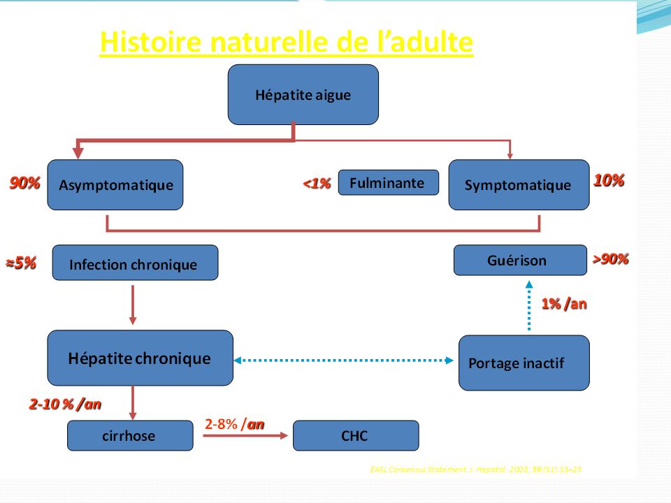
INTRODUCTION (II) Histoire naturelle Infection HCV Chronicité
60% à 85% Guérison 15% à 40% Cirrhose 10% à 15% Stable 85% à 90% This slide illustrates the natural history of hepatitis C infection. After acute infection, 15% to 40% of HCV-infected individuals appear to spontaneously clear the virus within approximately 6 months, thereby resolving their infection without sequel. Although their hepatitis C antibody test will remain positive and confirm the presence of prior infection, sensitive molecular tests will not show the presence of circulating HCV, and therefore, the patient is not at risk for developing hepatitis C–mediated liver disease or transmitting the disease to others. However, 60% to 85% of exposed individuals will not clear the virus and therefore will remain chronically infected. The most important consequence of chronic HCV infection is progressive liver fibrosis leading to cirrhosis, end-stage liver disease, and hepatocellular carcinoma (HCC). Fortunately, only approximately 10% to 15% of patients with HCV infection progress to cirrhosis over a period of 20 years after infection. One of the most serious complications of cirrhosis is the development of liver cancer (HCC) and liver failure. Hepatocellular carcinoma occurs almost exclusively in patients who have already progressed to cirrhosis. It has been estimated that up to 3% of people with cirrhosis develop HCC each year, and approximately 25% of patients with cirrhosis can be expected to develop HCC or liver failure. Thus, liver cancer and liver failure occur in approximately 2% to 4% of patients who are exposed to HCV over a 20- to 25-year period. Progression lente 75% Complications 25% CHC (2% - 4%) NIH Management of Hepatitis C Consensus Conference Statement. June Accessed April 2007. 22
. Fortunately, only approximately 10% to 15% of patients with HCV infection progress to cirrhosis over a period of 20 years after infection. One of the most serious complications of cirrhosis is the development of liver cancer (HCC) and liver failure. Hepatocellular carcinoma occurs almost exclusively in patients who have already progressed to cirrhosis. It has been estimated that up to 3% of people with cirrhosis develop HCC each year, and approximately 25% of patients with cirrhosis can be expected to develop HCC or liver failure. Thus, liver cancer and liver failure occur in approximately 2% to 4% of patients who are exposed to HCV over a 20- to 25-year period. Progression lente. 75% Complications. 25% CHC (2% - 4%) NIH Management of Hepatitis C Consensus Conference Statement. June Accessed April")
23
Circonstances de découverte d’une sérologie VHC (+)
- Dépistage ciblé: Profession exposée, VIH +, hémodialysés, cas familial … - Dépistage systématique: Don de sang, Femme enceinte, Chimio., Bilan Prénuptial Symptomatiques 37% Cirrhose 7% When patients with hepatitis C are asked how they feel, the majority—approximately 56%—will say they are fine and have no discernable symptoms to report. Only approximately 37% describe symptoms, the most common of which is fatigue, which is reported by 80% of patients. An additional 7% have more advanced liver disease, or cirrhosis, and may have detectable changes on the physical exam. Therefore, screening all of your patients for HCV infection is very important. Please see the worksheets and pocket guides related to screening and diagnosis in an injection drug user population found in this binder and online at 56% Asymptomatiques Hepatitis Program, 1995. 23

24
-Ac anti-HCV (sujets à risque ou don de sang) ARN viral+
- lésions Histo. Caractéristiques mais non pathognomoniques: agrégats ou follicules lymphoïdes; 44 à 78 %, Altérations épithélium des canaux biliaires sans ductopénie Stéatose: macrovacuolaire, minime à modérée, Lésions de nécrose parcellaire modérées voire discrètes.

25
Anamnèse Examen clinique
Facteurs de risque de contamination virale Présence et la chronicité des symptômes: - Ictère - Asthénie +++ Co-morbidité(s) connue(s) Facteurs aggravants: Ethylisme Signes d’hépatopathie Complications: ascite, EH Surpoids: BMI+++ Examen somatique complet Manif. extra- hépatiques: cutanées, thyroïdiens…
 connue(s) Facteurs aggravants: Ethylisme. Signes d’hépatopathie. Complications: ascite, EH. Surpoids: BMI+++ Examen somatique complet. Manif. extra- hépatiques: cutanées, thyroïdiens…")
26
Bilan biologique Bilan morphologique
Bilan hépatique complet: - Bilirubine totale+ conjuguée - ALAT / ASAT - PAL / GGT - TP / Albuminémie NFS: cytopénie:hypersplénisme? Manifestations extra-hépatiques: - Cryoglobulinémie - Bilan rénal+chimie des urines (glomérulonéphrite) Échographie abdominale: - Anomalies du parenchyme hépatique, signes HTP - Nodule suspect FOGD (si HTP): VO-VG-gastropathie?
 Échographie abdominale: - Anomalies du parenchyme hépatique, signes HTP. - Nodule suspect. FOGD (si HTP): VO-VG-gastropathie")
27
Les Ac anti-VHC Y Y Y Y Y Y HCV infecte les hépatocytes
Protéines HCV exprimées à la surface des hépatocytes Production d’Ac par l’hôte Détectables à partir 2-6 mois (fenêtre sérologique) Y Infection with HCV is a leading cause of chronic liver disease worldwide. The World Health Organization estimates that approximately 170 million individuals, or 3% of the world population, are infected by HCV. Hepatitis C virus is the most common blood-borne infection in the United States. Hepatitis C virus transmission occurs primarily through exposure to infected blood. This slide illustrates the process of hepatitis C infection and antibody production that occurs after a person has been exposed to HCV. The virus infects a liver cell, called a hepatocyte, and releases its genetic material, or RNA. This RNA causes the host cell to make large quantities of viral proteins, or antigens, that are then expressed on the surface of the infected hepatocyte. The host’s immune system recognizes these viral antigens as being foreign and reacts by producing antibodies to them (represented by the Ys), which bind to the infected cell. Interestingly, HCV does not directly damage the liver because it does not kill the cells. Instead, it causes the host’s own immune system to attack the liver, which can result in inflammation and the formation of scar tissue. It is also important to recognize that the antibodies that develop in response to infection with HCV do not lead to immunity to reinfection, as is commonly seen with other viral infections. Instead, antibodies to HCV are simply markers of prior exposure, like footprints. If an individual is HCV antibody positive but virus negative, and then participates in risky behaviors, he or she still has the potential to become reinfected with HCV. Y Y Y Y Y Y Y Y 27
 Y. Infection with HCV is a leading cause of chronic liver disease worldwide. The World Health Organization estimates that approximately 170 million individuals, or 3% of the world population, are infected by HCV. Hepatitis C virus is the most common blood-borne infection in the United States. Hepatitis C virus transmission occurs primarily through exposure to infected blood. This slide illustrates the process of hepatitis C infection and antibody production that occurs after a person has been exposed to HCV. The virus infects a liver cell, called a hepatocyte, and releases its genetic material, or RNA. This RNA causes the host cell to make large quantities of viral proteins, or antigens, that are then expressed on the surface of the infected hepatocyte. The host’s immune system recognizes these viral antigens as being foreign and reacts by producing antibodies to them (represented by the Ys), which bind to the infected cell. Interestingly, HCV does not directly damage the liver because it does not kill the cells. Instead, it causes the host’s own immune system to attack the liver, which can result in inflammation and the formation of scar tissue. It is also important to recognize that the antibodies that develop in response to infection with HCV do not lead to immunity to reinfection, as is commonly seen with other viral infections. Instead, antibodies to HCV are simply markers of prior exposure, like footprints. If an individual is HCV antibody positive but virus negative, and then participates in risky behaviors, he or she still has the potential to become reinfected with HCV. Y. Y. Y. Y. Y. Y. Y. Y. 27.")
28
Détection des AC anti VHC
Test ImmunoEnzymatique ELISA 3ème génération: - Sensibilité : 97% - 100% - Spécificité: 99% Faux négatifs: - Etats d’immunosuppression - IRC hémodialysés - Co-infection VIH - Transplantation Faux positifs: Rares Affections auto-immunes The most common anti-HCV antibody tests are enzyme-linked immunosorbent assay (ELISA) tests. These tests use a small plastic plate impregnated with HCV proteins, graphically represented as triangles. The patient’s serum is then added to the plate, and if it contains any anti-HCV antibodies, they will bind to the HCV proteins. Next, a second tagged antibody that will detect any anti-HCV that is bound to the plate is added, and a positive reaction is detected by the marker changing color. The ELISA antibody tests are highly sensitive; the positive predictive value of ELISA tests is 95% in subjects who have HCV risk factors and elevated ALT. However, false-positive ELISA results sometimes occur, such as in patients with autoimmune conditions. A positive test result in a person who is at low risk for HCV infection is potentially a false positive and a virologic test (for HCV RNA) is necessary to confirm a diagnosis of chronic hepatitis C. A negative ELISA test result is sufficient to exclude a diagnosis of hepatitis C in immunocompetent individuals. However, immunosuppressed individuals (who have difficulty producing antibodies), including transplantation recipients, chronic renal failure patients on dialysis, or HIV-coinfected patients, may have false-negative ELISA results. Again, if immunosuppression is suspected, a virologic assay is necessary. NIH Management of Hepatitis C Consensus Conference Statement. June 2002. Accessed April 10, 2007. Carithers RL Jr, et al. Semin Liver Dis. 2000;159 Pawlosky JM. Hepatology. 2002;36(suppl 1):S65-S73. 28
 tests. These tests use a small plastic plate impregnated with HCV proteins, graphically represented as triangles. The patient’s serum is then added to the plate, and if it contains any anti-HCV antibodies, they will bind to the HCV proteins. Next, a second tagged antibody that will detect any anti-HCV that is bound to the plate is added, and a positive reaction is detected by the marker changing color. The ELISA antibody tests are highly sensitive; the positive predictive value of ELISA tests is 95% in subjects who have HCV risk factors and elevated ALT. However, false-positive ELISA results sometimes occur, such as in patients with autoimmune conditions. A positive test result in a person who is at low risk for HCV infection is potentially a false positive and a virologic test (for HCV RNA) is necessary to confirm a diagnosis of chronic hepatitis C. A negative ELISA test result is sufficient to exclude a diagnosis of hepatitis C in immunocompetent individuals. However, immunosuppressed individuals (who have difficulty producing antibodies), including transplantation recipients, chronic renal failure patients on dialysis, or HIV-coinfected patients, may have false-negative ELISA results. Again, if immunosuppression is suspected, a virologic assay is necessary. NIH Management of Hepatitis C Consensus Conference Statement. June Accessed April 10, Carithers RL Jr, et al. Semin Liver Dis. 2000;159. Pawlosky JM. Hepatology. 2002;36(suppl 1):S65-S")
29
Charge virale = PCR La quantification de l’ARN-VHC par PCR “temps réel”

30
Anti HCV+ Test virologique de confirmation PCR -Infection guérie Infection persistante -Faux +

31
Cinétique des marqueurs virologiques au cours d’une hépatite chronique

32
-TRT: PEGASYS® + Ribavirine

33
2-Hépatites médicamenteuse:
-Traitement prolongé (au moins 6 mois) entrainant une Nécrose hépatocytaire = HC, voire cirrhose. -Principaux médicaments responsables: l’acide acétylsalicylique,Amineptine, chlorydrate de papavérine, dantrolène, Halothane, Isoniazide, Méthyldopa, Nitrofurontoïne, Paracétamol, phénylbutazone, Prophylthiouracil, Sulfamides, acétaminophene, clométacine, diclofénac, glifanan, oxyphénisatine, etc.
 entrainant une Nécrose hépatocytaire = HC, voire cirrhose. -Principaux médicaments responsables: l’acide acétylsalicylique,Amineptine, chlorydrate de papavérine, dantrolène, Halothane, Isoniazide, Méthyldopa, Nitrofurontoïne, Paracétamol, phénylbutazone, Prophylthiouracil, Sulfamides, acétaminophene, clométacine, diclofénac, glifanan, oxyphénisatine, etc.")
34
-Mécanisme: effet toxique direct ou de métabolite (s) ou par une réponse immuno-allergique.
 ou par une réponse immuno-allergique.")
35
3-MALADIE DE WILSON : -liée à un désordre du métabolisme du cuivre, autosomale récessive se manifestant par une hépatite fulminante ou hépatite chronique et cirrhose. -Le gène défectif (ATP7B) se localise dans le long bras du chromosome 13. -La mutation commune, la H1069Q altère la fonction de transport protéique du cuivre. -A considérer chez les sujets jeunes. -histopathologie: stéatose,, accumulation de cuivre, corps de Mallory.
 se localise dans le long bras du chromosome 13. -La mutation commune, la H1069Q altère la fonction de transport protéique du cuivre. -A considérer chez les sujets jeunes. -histopathologie: stéatose,, accumulation de cuivre, corps de Mallory.")
36
5-Hépatite auto-immune :
- D’étiologie inconnue -Caractérisée par: -Piece meal necrosis -Hypergammaglobiline -Positivité des Auto-anticorps -Diagnostic : Marqueurs Viraux négatifs Absence d’exposition au sang, éthanol, drogues -Globulines 1,5 x LSN Auto-Ac > 1/80 (ANA, SMA, p-ANCA, ASPGR, LKM)
")
37
infiltrat plasmatique, PMN, nécrose en pont ou multiacinaire)
Rechercher: thyroïdite, PR, CBP, CSP, Diabète, MICI, M coeliaque (Ac endomysium, transglutaminase, gliadines) Type 1: ANA et AML au moins > 1/80 chez l’adulte, Ac anti actine les plus spécifiques, SLA/LP ++ mais dans 10%, atypiques pANCA, Type 2: Anti–LKM-1, anti–LC-1 -TRT:CTC+immunosuppresseur
 Type 1: ANA et AML au moins > 1/80 chez l’adulte, Ac anti actine les plus spécifiques, SLA/LP ++ mais dans 10%, atypiques pANCA, Type 2: Anti–LKM-1, anti–LC-1. -TRT:CTC+immunosuppresseur.")
Présentations similaires